Home / About / Center Grants /
 The University of Washington’s Eunice Kennedy Shriver Intellectual and Developmental Disabilities Research Center (IDDRC), based at the Institute on Human Development and Disability (IHDD), provides a comprehensive interdisciplinary research program in the field of intellectual and developmental disabilities (IDD).
The IDDRC empowers research affiliates by providing access to cutting edge technologies and data analysis techniques.
Core services provided include experimental design support, grant development, equipment and space access, staff training, research expertise and best practices, and the coordination of UW resources. We also provide support for early career researchers. Scientific core support facilities are as follows: (1) Animal Behavior, (2) Brain Imaging, (3) Clinical Translational, and (4) Genetics.
IDDRC Director

Our Core Facilities include:

Animal Behavior Core
The Animal Behavior Core provides multi-pronged services for Research Affiliates using preclinical animal models of developmental disorders.

Brain Imaging Core
The overall goal of the Brain Imaging Core is to support affiliates and train junior investigators to perform scientifically sound research by providing…

Clinical Translational Core
The Clinical Translational Core functions as a service to IDDRC scientists to organize, integrate, and promote the use of translational research resources.

Genetics Core
The Genetics Core is designed to integrate cutting-edge genomic technologies into the research programs of investigators to enhance the understanding of IDD..

Research Affiliate Program
The University of Washington IDDRC’s Research Affiliate program fosters collaboration among scientists who conduct research in the field of intellectual and developmental disabilities.
IDDRC Director
Don’t forget to cite the grant 5P50HD103524 to credit the IDDRC for the support you have received!
Our Collaborative Research Areas include:
 The purpose of the Adults and Elders Program is to promote the health and overall quality of life of adults with developmental disabilities. This values-based program emphasizes an individual’s inclusion, self-determination, productivity, and independence. Read more…
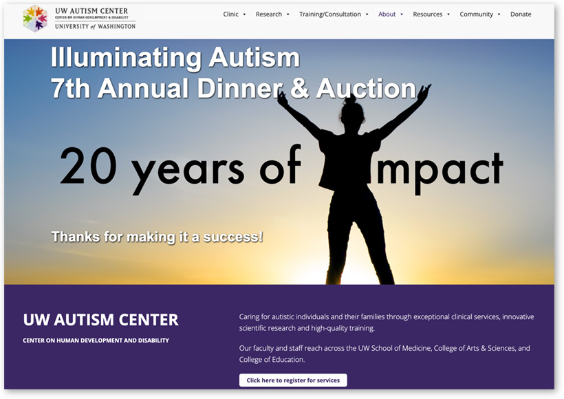 The UW Autism Center (UWAC) was founded in 2000 and is one of the first autism centers in the country to provide clinical services, professional training, and internationally recognized research under one roof. Clinically, UWAC provides intervention services, diagnostic evaluations, and program consultation for children from birth through young adulthood with Autism Spectrum Disorders. Read more…
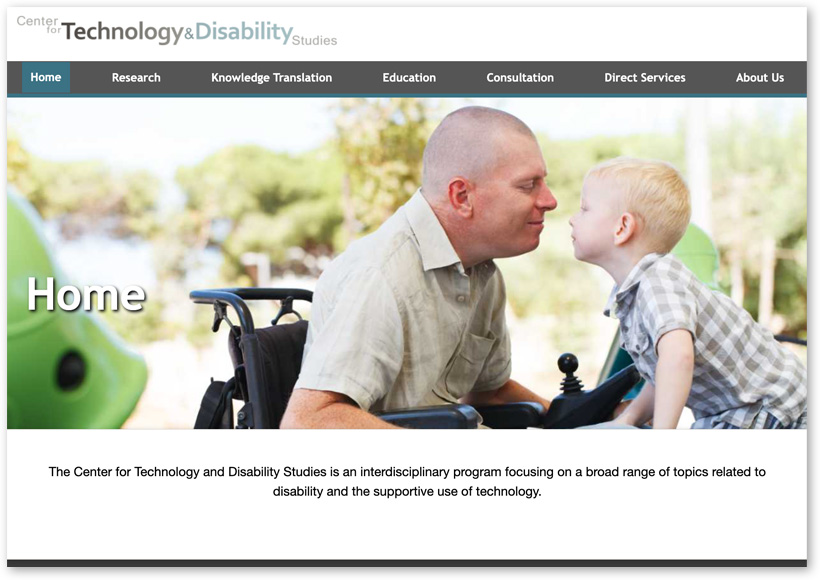 The Center for Technology and Disability Studies(CTDS) works to advance assistive technology (AT) and accessible information systems to support individuals with disabilities in accessing opportunities in education, their community, and employment. Read more…
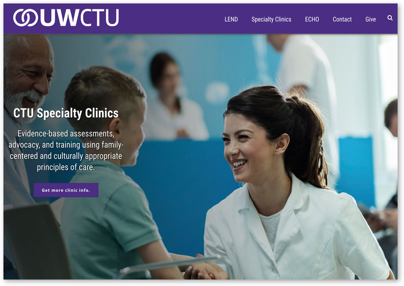 The Clinical Training Unit (CTU) is an interdisciplinary program that provides training, research, and exemplary services in the assessment and treatment of children with or at risk for developmental disabilities, using a family-centered, community-based, culturally competent approach. Read more…
 The Genetics Program comprises clinics that serve individuals at risk for or with a genetic disorder or disability. Clinics offer diagnosis, assessment, treatment, and counseling services to meet the needs of these clients. Read more…
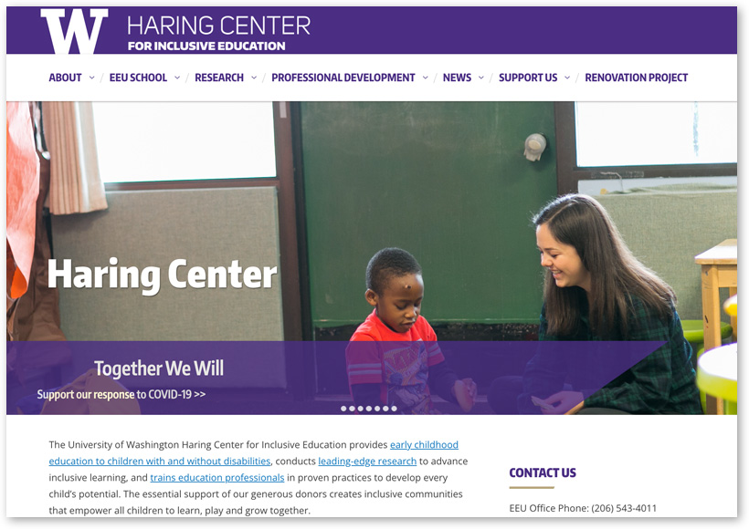 The Norris and Dorothy Haring Center for Research and Training in Inclusive Education provides early childhood education to children with and without disabilities, conducts leading-edge research to advance inclusive learning, and champions the best practices we develop beyond our walls so that every child can reach their full potential. Read more…
The Norris and Dorothy Haring Center for Research and Training in Inclusive Education provides early childhood education to children with and without disabilities, conducts leading-edge research to advance inclusive learning, and champions the best practices we develop beyond our walls so that every child can reach their full potential. Read more…
The Norris and Dorothy Haring Center for Research and Training in Inclusive Education provides early childhood education to children with and without disabilities, conducts leading-edge research to advance inclusive learning, and champions the best practices we develop beyond our walls so that every child can reach their full potential. Read more…