Exploring and Stretching the Boundaries of Brain Imaging
by Joel Schwarz
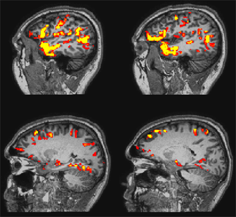
Tom Grabowski and other researchers affiliated with CHDD use Magnetic Resonance Imaging (MRI) to study brain activation in patients with epilepsy. Here the orange pixels show brain activity in the left temporal lobe of a normal patient who is being tested for comprehension of stories that are delivered by headphones in the MRI scanner.
University of Washington researchers are exploring new brain imaging techniques that someday may enable clinicians to better understand and treat patients suffering from such conditions as stroke, epilepsy, Alzheimer's disease and other forms of dementia. "We are trying to develop imaging biomarkers of prognosis and responsiveness to treatment. This may enable us to predict whether or not people will respond to treatment," said Thomas Grabowski, Jr., M.D., director of the Integrated Brain Imaging Center (IBIC) and a research affiliate of the Center on Human Development and Disability (CHDD).
As director of IBIC, Grabowski, who is also a professor of radiology and neurology, is spearheading an effort to promote interdisciplinary collaboration in human neuroimaging, cognitive neuroscience and medicine across the UW campus. In addition, he has an ambitious research program of his own focusing on the neural basis of language processes and language disorders, and how the left temporal lobe is organized.
This fall has been a particularly busy time for Grabowski because IBIC received a three-year, $3 million federal grant that will promote the formation of new multi-disciplinary teams. The grant also brings together half a dozen centers and adds imaging components to their work. "We are pushing the envelope methodologically in how functional magnetic resonance imaging (fMRI) works and trying to focus interdisciplinary expertise in one center," he said. With the new grant, for example, IBIC will join with CHDD and the Northwest Epilepsy Center in its on-going research into temporal lobe epilepsy. "What happens now in treating epilepsy is that we succeed with patients but sometimes they end up with a language problem, such as an inability to retrieve names. We think we can use imaging to see who might end up with language problems."
Another IBIC project will focus on the posterior cingulate, a brain region that is called "ground zero" for Alzheimer's disease, to use magnetic resonance spectroscopy to look for a noninvasive biomarker for the condition and to develop tools to chart brain changes. Funding also will support imaging research in the on-going Seattle Longitudinal Study which has discovered that there is a small subset of people whose memories in mid-life improve with age. Researchers want to know if this condition is stable and what distinguishes these individuals from other people. Grabowski said the grant also will enable researchers to study how brain regions react with each other and to early aging and epilepsy, as well as how to combine electrophysiology techniques with imaging.
His own research looks at how the brain supports language, especially how the left hemisphere retrieves words. "My interest is in the processes involved in retrieving words and how these processes are affected by stroke, epilepsy, and age," said Grabowski. "People with damage to the left temporal lobe can develop selective trouble in retrieving the specific names of things. This kind of retrieval problem is also characteristic of Alzheimer's. We are trying to extract information from images of a patient's brain as it is working on a task. We might ask patients to tell us the name of each of a series of objects they are shown, some common and not common. This work is important because we may be able to use imaging to understand how language trouble relates to the progression of a disease and we might be able to use it to predict which people will suffer language impairment from surgery, epilepsy or Alzheimer's."
Grabowski is using an approach he calls "systems level neuroimaging" and believes it has enormous potential. "It can provide a big difference comparable to being able to see that the lights are on in a room versus being able to see who is talking to whom in that room. In the future, we envision using it to be able to see where language is supported and where epilepsy is focused in an individual, and to be able to understand how to operate more precisely on that patient. Currently, trying to detect this is quite invasive and a burden on the patient. "This kind of imaging is a smarter way of understanding what the signals from the brain mean."
|