Genetics 371B, Autumn 2000 Answer key, exam 2: 17 November |
Answer questions 1-4 and EITHER #5 OR #6, but not both.
Answers are in blue
1. | A certain X-linked dominant disease in humans occurs when there are more than 36 repeats of a CAG trinucleotide in a particular gene. The unique sequence flanking the repeat region is as follows:
|
|
(a) | Which of the primer(s) from the list below would you would use, if you wanted to PCR-amplify the entire trinculeotide repeat region? (4 pts)
Primer: #1) 5'-TCTCCTAGTGGTCATGGGTC-3' #2) 5'-AGAGGATCACCAGTACCCAG-3' <--- this one... #3) 5'-GTCCAGACTCACGTGTTGGA-3' <--- and this one #4) 5'-TCCAACACGTGAGTCTGGAC-3' #5) 5'-(CAG)7-3' |
|
(b) |
Shown below is the outline of a pedigree for the disease described above, and a representation of a gel showing PCR-amplified fragments detecting the number of CAG repeats. The DNA corresponding to each individual is directly below his or her place in the pedigree. Based on the information you have been given, fill in the pedigree to show the sex and phenotype (affected vs. unaffected) of all individuals except II-9 (save that one for the next question). (10 pts) |
|
(c) | What is unusual about individual II-9? Suggest two mechanisms that could explain the genotype of II-9, stating whether that individual would be phenotypically female or male in each case. (6 pts)
The individual appears to have just a single copy of the polymorphic locus, so we'd predict it was an XY male -- except that the allele appears to be derived from the father (26 repeats), and the father should be contributing only a Y to his son. Several possible explanations:
|
2. | (a) | A mutation in a bacterial DNA polymerase results in an elevated rate of nucleotide mis-incorporation during DNA replication. Below is a portion of a sequence of newly replicated DNA from this mutant bacterium. "Me" indicates a methyl group on the adenine. (8 pts)
|
||||||||||||
(b) | State whether each of the following mutations would result in dominant , recessive , co-dominant , or incompletely dominant phenotypes for the mutant alleles. Explain BRIEFLY. (6 pts)
|
|||||||||||||
(c) | BRIEFLY explain how a mutation in a tumor suppressor gene can show dominant inheritance in pedigrees and yet be recessive for cancer formation at the cellular level. (6 pts)
A heterozygous cell still has the wild type phenotype, but since it takes only one more mutation or a mitotic recombination event in any one dividing cell in the tissue, there is a very high probability that at least one cell will acquire that second change. Therefore, although the mutant allele is recessive, at the whole-person level, the disease trait is inherited in dominant fashion (perhaps with reduced penetrance). |
3. | ![]() The fish is shiny and green with broad scales, as expected, but you notice occasional spots of scales that look different. |
|
(a) | What are the two most common types of spots you expect to see in this fish? In what proportions would you expect to see these spots? [HINT: You should be able to answer this part just by looking at the diagram above, before you draw the chromosome diagrams for part (b).] (6 pts)
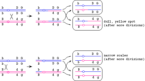
Most common: dull, yellow (mitotic recombination with single crossover between the centromere and D/d -- that is the largest interval in the map) 2nd-most common: narrow scales (single cross between the centromere and B/b -- that is the 2nd largest interval in the map) The proportions: 3 dull, yellow : 2 narrow (because that's the relative proportions of the two intervals that each have a single crossover). |
|
(b) | Draw chromosomal diagrams explaining the formation of one of the two spots you have listed in (a). (Your choice of which spot to diagram, but you must state clearly which one you are doing.) (10 pts)
|
|
(c) |
What is the least common type of lone spot you expect to see in this fish? In one sentence, say what kind of event would give rise to this type of spot. (No need for diagrams.) (4 pts) A lone spot of dull scales...a result of a double (mitotic) recombination, one between the centromere and D/d, one between D/d and G/g. [Note: a triple crossover would be even more rare, but it wouldn't give lone spots.] |
4. | Males of the worm C. elegans are XO they have one X chromosome and no Y chromosome, and their sperm have either one X or no X chromosome. You have identified two autosomal recessive mutations: nd-1 causes X chromosome non-disjunction in every cell undergoing meiosis I, and nd-2 causes X chromosome nondisjunction in every cell undergoing meiosis II. For all three questions below, assume that the eggs came from wild type XX animals. | |
(a) |
What fraction of a +/nd-1 male worm's progeny do you expect will be male (XO)? Explain BRIEFLY. (4 pts)
|
|
(b) |
What fraction of an nd-1/nd-1 male worm's progeny will be male (XO) ? Explain BRIEFLY. (6 pts) 0.5-- same as with (a), because meiosis-I non-disjunction does not affect an XO animal (there is no homolog to disjoin from in the first place). |
|
(c) | What fraction of an nd-2/nd-2 male worm's gametes can produce offspring with normal chromosome numbers? What will be the sex of these offspring? Explain BRIEFLY. (10 pts)
NOTE: For the first two parts, we are assuming that XXX animals are viable. |
Answer Question 5 or Question 6, BUT NOT BOTH. If you answer both, we will not grade #6.
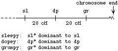
5. | You are working on 3 behavioral genes on the X chromosome of Drosophila :
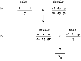
Wild type males are crossed to completely recessive females. From the progeny of this cross, single females are picked and mated to recessive males, to produce an F2 generation: |
|||||||||||||||||||||
(a) | Now imagine that the parental wild type male was treated with X-rays, such that his X chromosome acquired a deletion that removed the grumpy locus. In one or two sentences, describe how this deletion would affect the F2 progeny ratios compared to an unirradiated control. (6 pts)
Because the deletion takes out sequences to the right of the dopey locus, we will not see recombination in the dp - gr interval. The relative proportion of parental types in the progeny will increase, and the relative proportion of recombinants will decrease. |
|||||||||||||||||||||
(b) |
Predict the F2 progeny phenotypes and numbers for this deletion strain assuming that you counted a total of 1000 progeny. (14 pts) The F1 female here is:
so the gametes and progeny are going to be:
(Looking at just the female progeny. The male prpgeny will show the same phenotypes, assuming that they are not dead because of the deletion.) Note: If you hypothesized that the deletion breakpoint was just to the left of gr, you could postulate that recombinants in the dp-gr interval would be recovered also, although at reduced frequency. That answer will also be accepted, provided that you state your hypothesis and the predictions of that hypothesis clearly. |
Answer Question 5 or Question 6, BUT NOT BOTH. If you answer both, we will not grade #6.
6. | The map below represents a 200 kb (kilo-base pair) portion of the mouse X chromosome; SfiI restriction enzyme sites are marked ("SI"). Various deletions (i though iv) found in this portion of the chromosome are shown. The gaps in the bars represent the portions of the DNA that are missing in the deletions, but remember that the breakpoints of the deletion are joined together.
|
|
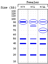
(a) | DNA was obtained from various mice (or aborted mouse fetuses), cut with SfiI , and size-separated by gel electrophoresis. For each of the strains (listed above the outline of the gel below), mark the locations of the bands you expect for this segment of DNA. Then circle those bands that will hybridize to the probe shown as a cross-hatched bar in the map. (12 pts)
|
|
(b) |
For some of these deletions (indicated to the right of the map), it was found that female fetuses heterozygous for the deletion died before birth; cell of these fetuses lacked Barr bodies. Where in this segment of DNA is the critical portion of the X inactivation center located? (Give the coordinates in kb from the left end of the map.) Speculate BRIEFLY on why deletions i, iii and iv result in absence of Barr bodies and and in death of the fetus. (8 pts) The absence of Barr bodies in some deletion strains suggests that those strains lack the X inactivation center -- the X chromosome without the XIC is not counted as an X, so neither X chromosome is inactivated. The resulting imbalance in X chromosome genes presumably causes the lethality of the fetus. The deleted portion in common in all the strains that had aborted female fetuses was 110-120 kb from the left end of the map -- so that must be the location of the X-inactivatin center. |