Answer key, practice problems 2000--week 6

1. (a)

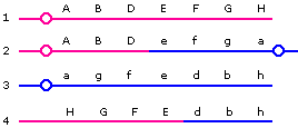
Products 1 and 3 will be viable. Product 2 is dicentric, has two copies of gene A and lacks gene H; product 4 has two copies of gene H, and lacks gene A and a centromere. Therefore, these two products will probably fail to form viable progeny.
(b)

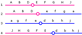
Again, Products 1 and 3 will be viable, while 2 and 4 (having duplications of certain segments and lacking other segments) will each probably be inviable. (Note that the products depicted here are those that would result from a crossover between the E locus and the centromere; the crossover could just as well occur between the D locus and the centromere.)

[See Lecture 17 for details on how these results are obtained.]
2. The observation can be explained as a case of sex reversal, where a piece of the Y chromosome (carrying the SRY gene) is translocated to another chromosome (either an autosome or an X) such that an XX individual nevertheless develops male characteristics because of the translocated portion.

3. (a) The 38-year old has a higher risk of a Down syndrome baby, because the probability of nondisjunction during meiosis increases with age in human females.
(b) The family history of Down syndrome suggests that this may be a case of translocation Down syndrome–in which case, the younger woman (belonging to that family) has a higher risk of a Down syndrome baby (because the chance of nondisjunction in a 38-year old woman is about 1 in 100 – see pg 69 of the lecture notes – while the chance of translocation Down carrier having a Down baby is 1 in 4).

4. (a) The mother must have been heterozygous G/g, while the father was hemizygous normal G/(Y). Colorblind Turner syndrome (XO) females must have resulted from fertilization of a g egg by a sperm lacking a sex chromosome; nondisjunction could have occurred at meiosis I or meiosis II in the father. Colorblind Klinefelter (XXY) males must have resulted from fertilization of gg eggs by normal Y-bearing sperm; nondisjunction occurred in meiosis II in the mother.
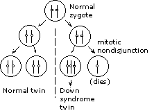
(b) Identical (monozygotic) twins arise when an early embryo splits so that each portion develops into an individual fetus. In this instance, the zygote must have been normal. The split occurred at the two-cell stage; one of the resulting cells divided normally, giving the normal twin, while the other cell had a mitotic nondisjunction, giving the Down syndrome twin.

5.

Six types of sperm; if the two Y chromosomes are labeled Y1 and Y2 to distinguish them from each other, the gamete types are:

XY1
Y2
XY2
Y1
Y1Y2
X

in the ratio 1:1:1:1:2:2.

6. (a) Because the plant height and color genes are on separate chromosomes, they should assort independently; the cross should give TD, Td, tD, and td progeny in 1:1:1:1 ratio. Instead, only the parental phenotypes (TD and td) are seen.
(b)

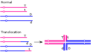
The absence of the non-parental types and the semi-sterility suggest that the explanation may be a translocation. One possible configuration is shown:

The "adjacent" pattern of segregation would give Tt and Dd gametes, while the "alternate" pattern would give TD and td. The Tt and Dd gametes would be inviable, so the only viable progeny would have TD and td phenotypes–the parental types.

Note that there is more than one configuration that would fit the results. For example, the t and d alleles need not be on the translocated segment.

7. (a) The F1 females should be heterozygous at all loci. We would normally expect to see recombination in each interval, giving up to 26 = 64 different progeny phenotypes (in a ratio that would depend on the map distances). The presence of only four progeny phenotypes is not normally to be expected. (One could postulate that pairs of loci are very tightly linked, but that does not explain the lack of recombinants between the ends of the group.)
(b) A deletion could be ruled out because half the F2 males would inherit an X chromosome lacking genes, and would probably fail to develop. Translocations are also unlikely to give the observed results, because the phenotype is a reduction in observed recombinant progeny (translocations cause semisterility, but there is no reason not to expect recombinants (draw it out and confirm it for yourself).
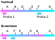
(c) The lack of recombination between A and B and between F and G suggests that the entire portion between A and G (i.e., B through F) is inverted.
(d)
A variety of molecular tests is possible. For example, if the inversion is as predicted, one can set up Southern blots, using probes for the presumptive junction regions. In the example shown here, Probes 1 and 2 will hybridize to different restriction fragments (of different sizes) if the chromosome is normal. In contrast, if the inversion is as shown, probes 1 and 2 will both hybridize to the same fragment. [Restriction enzyme sites are depicted as vertical bars. Knowing the restriction map for the whole chromosome, we can pick a restriction enzyme that has suitably located sites as shown.]
(e) Single crossovers are not expected to give viable recombinant gametes. However, rare double crossovers can be viable. In this instance, a double crossover -- one crossover between B and D loci and one between E and F -- would give the observed result.

8.

If the two T-allele bearing homologs are called T1 and T2, and the two t-allele bearing homologs are t1 and t2, there are three possible sets of pairings, giving the gametes shown:

Pairing Gamete genotypes
T1T2 and t1t2 (i.e., T1 homolog paired to T2 homolog, etc.) T1t1, T2t2, T1t2, T2t1
T1t1 and T2t2 T1T2, t1t2, T1t2, T2t1
T1t2 and T2t1 T1T2, t1t2, T1t1, T2t2

Thus, the gamete genotypes are TT, Tt and tt in 1:4:1 ratio, or 5:1 ratio of T_:tt. If mated to tttt plants (whose gametes will all be tt), the progeny are expected to be T___:tttt (i.e., tall and short) in 5:1 ratio.

9. (a)

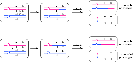
The patches probably arose by mitotic recombination. Recombination between the two loci would give lone spots of the recessive phenotype of the more centromere-distal locus, while recombination between the centromere and the two loci would give twin spots. From this logic, we can conclude that the rd locus must be closer to the centromere than the b locus. (An alternative explanation for the lone spots is mitotic nondisjunction, but that wouldn't explain the twin spots.)

(b)

Because the twin spots and lone spots occurred in 6:5 ratio, the centromere-rd distance and the rd-b distance must be in the approximate ratio of 6:5:

(c)

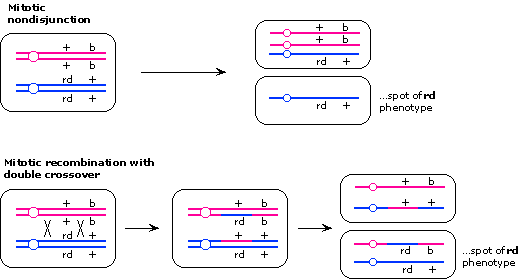
Lone spots of rd phenotype could arise either from mitotic nondisjunction or by mitotic recombination with double crossover, one crossover between the centromere and rd and one crossover between rd and b.

10.
The most distal markers must be y and g. Because yellow and rough are seen in twin spots with each other, but not with mottled or sparse, genes y and r must lie on one arm of the chromosome. Likewise, m and g must lie on the other arm of the chromosome. Therefore, the gene order on the chromosome is:
y       r                          centromere  m             g
|-------|-------------------------------O------|-------------|
    7               32                      6         12

11. (a) Sectors of different sizes will arise depending on when during growth of the colony the mitotic recombination event occurred--the earlier the recombination, the larger the sector. Half-sectored colonies reflect mitotic recombination events that occurred in the first division of the cell that eventually formed the colony.
(b)

The problem in measuring recombination frequency is estimating how many cell divisions have occurred. However, we do know how many cells underwent mitotic recombination to give sectors in the first division--it is the number of half-sectored colonies. We also know how many "first divisions" occurred--it is equal to the number of colonies on the plate. Therefore, the frequency of mitotic recombination = frequency of mitotic recombination in the first division = 2(# of half sectored colonies)/(total # of colonies).

[The frequency of half-sectored colonies has to be multiplied by 2 because half the time mitotic recombination occurs, it is expected to form double heterozygous daughter cells, which will not give a sector. If this is unclear, look at the Rb/rb example in p.81 of the lecture notes.]

12. (a) Because the only defect in the reduced-function allele is a point mutation, this portion of the chromosome will show the same gene order and map distances in affected individuals and unaffected individuals. With the elevated expression form, it is not possible to predict beforehand whether the chromosome is rearranged or not--and in fact, looking at the LOD scores, it is clear that the recombination distances are not the same as with the point mutation pedigrees. Therefore, to generate a map of the region in normal individuals, LOD scores based on the reduced-expression form of the disease should be used.
(b)
Either a deletion or an inversion in the G-PS2 interval would result in suppression of recombination in that interval, resulting in apparent tight linkage between G and PS2 and an apparent reduction in the PS1-PS2 map distance.
 
(c) The elevated expression phenotype is dominant, suggesting that there is an inappropriate activation of gene function--e.g., gene G is now juxtaposed with a promoter that causes inappropriate transcription of the gene, or the rearrangement results in a fusion protein that is resistant to the normal control mechanisms.