

Director: Ruikang (Ricky) Wang
Retina vessel quantification
Purpose
To quantifying the vasculature in macular OCTA images.
Input
En face projection of OCT-A scans from commercial and homebuilt OCT systems (.jpg/.png./tiff/.bmp)
User defined reginal masks (optional)
Adjustable segmentation parameters
Output
Vessel segmentation, binary skeleton and binary flow impairment zone map
Vessel area density, vessel diameter, vessel skeleton density and vessel complexity index of whole image and reginal masks
Validation
US patent US10354378B2
Chu, Z., Lin, J., Gao, C., Xin, C., Zhang, Q., Chen, C. L., ... & Wang, R. K. (2016). Quantitative assessment of the retinal microvasculature using optical coherence tomography angiography. Journal of biomedical optics, 21(6), 066008.
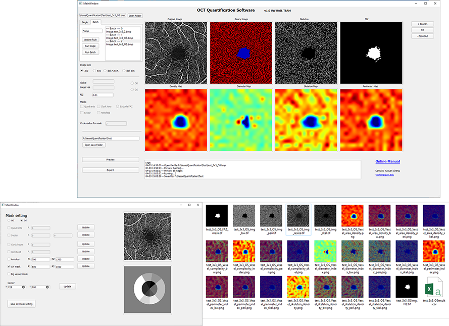
The retinal vessel quantification software
Displayed images:
Original OCTA image,
binary vessel map,
binary skeleton map,
and binary flow impairment zone map.
Vessel area density color map,
vessel diameter color map,
vessel skeleton density color map,
and vessel complexity index color map.
The user defined reginal mask tool..
The results of the software.
Full manual
Contact Info
Department of Bioengineering, N410 William H. Foege Building, 3720 15th Ave NE Seattle, WA 98195
E-mail: wangrk @ uw dot edu
Phone: (2o6) 616-5o25
