Picturing the Biochemistry of Disease
The Molecular Imaging Program at the UW is developing new methods to pictures the biochemistry of disease. These pictures are called PET (positron emission tomography) images. They use molecules labeled with radioisotopes with a short half-life to follow the rate of chemical reactions inside the body.
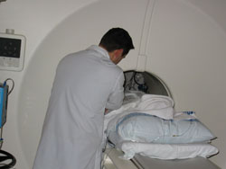 |
| PET Technologist Nathan Roybal helps a patient feel comfortable while in the scanner. |
The new tracers have been so useful in local studies that the Cancer Imaging Program of the National Cancer Institute is teaming up with GE Medical Systems for a multi-site research effort to evaluate several of the radio- pharmaceuticals pioneered at the UW. The first effort will involve clinical trials of 18F-fluorothymidine (FLT) as an imaging agent for positron emission tomography. One of the sites is UW Medical Center, with Dr. Janet Eary, professor of radiology, heading the clinical trial and Dr. Jeanne Link, associate professor of radiology, writing much of the Food and Drug Administration documentation for this new drug.
FLT shows promise for studying the growth of human cancers because it indicates how fast cells are multiplying. It mimics one of the nucleic acids and is used to determine the rate of DNA synthesis with non-invasive imaging. Cells take up FLT when making new DNA. Researchers can image the disease site and record the rate of DNA synthesis to measure cell growth in the tissues. The imaging radioisotope, Fluorine 18, has a half-life of 110 minutes.
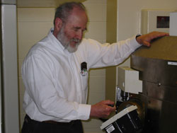 |
| Cyclotron operator Bob Murano checks for radiation with a geiger counter. |
"Because it has a short half life," said Dr. Kenneth Krohn, professor of radiology and director of the UW Radiochemistry Laboratory, " we can use this PET agent to image a patient today, the cancer doctor can treat the patient, and we can bring the patient back a within short time, such as a week later, for imaging to see if the treatment is working."
UW scientists in the Molecular Imaging Program in the Department of Radiology first made and tested the FTL imaging agent about six years ago. It is now used in research labs around the world. The new National Cancer Institute-sponsored imaging trials groups are performing multi-institutional studies to enable more widespread use of FLT.