I have had the opportunity to study a diversity of structures and critters – from teeth and body armor like tough scales, to softer tissues like lips and the discs of fishes that suck onto surfaces. Each project requires a set of imaging tools that reveal hard and soft anatomy in different ways. One of the great joys of my work is translating careful observations of anatomy into beautiful images and figures. 2D visualization like histology and photography let me see the world through clearly arranged points of view, while 3D imaging shows me context and relationships.
I started as a histologist, someone who slices things thinly to allow stains to reveal different tissues.
I use paraffin and plastic histology to measure qualitative and quantitative traits of tissues like how collagen is arranged or how much muscle is present to make predictions about their mechanics.
Pacus, the herbivorous cousins of notoriously carnivorous piranhas, have large multi-cuspid teeth that are hidden by large lips analogous to the tongues and snoots of giraffes and rhinos (Cohen and Kolmann, 2021). Using plastic histology, I sectioned the lips of the pacus to find that they are not muscular like those of mammals, but are instead composites with layers of collagen, fat and keratin (the material that makes up our fingernails, Figure 1). Sometimes it's important to not just see the morphology and arrangement of tissues but also know their identity. This is where paraffin histology comes into play. It revealed that some of the soft tissue in pacu lips is elastin: a protein responsible for stretchiness.

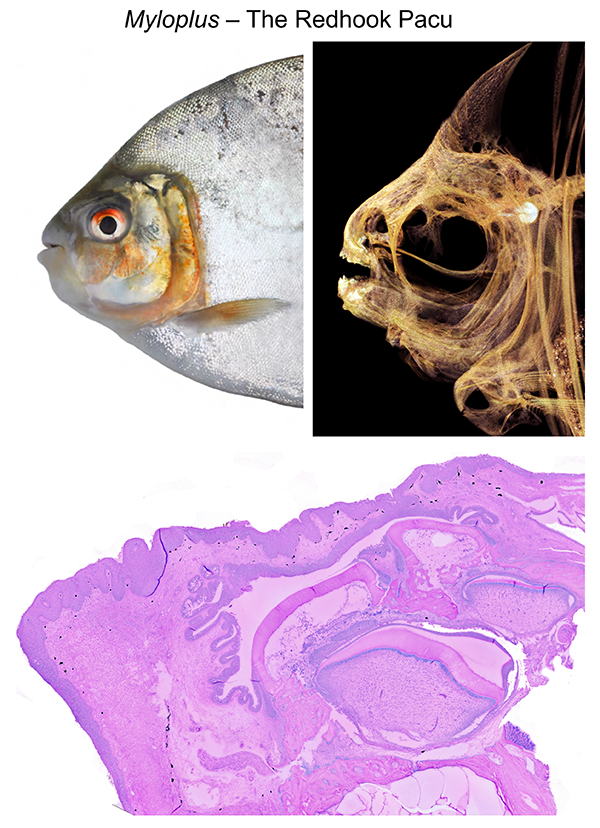 Fig. 1: Looking at soft stuff. Images of the Redhook pacu, Myloplus, showing what the fish looks like in life (adopted from Huie et al. 2019) vs. in CT. Below is a histology section through the lower lip revealing layers of collagen, keratin and other materials that help this species grab and hold onto food.
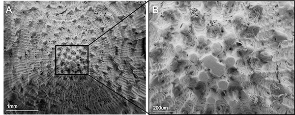
 Scanning electron microscopy (SEM) lets you see the outside topography of structures and has been crucial in seeing damage on spines, scales and teeth. Damage is the scar left behind when critters interact with food or the environment, and it shows how a given structure is being used. The Pacific spiny lumpsucker is a favorite fish of FHL researchers, and gets its name from the conical armor that covers its whole body (Figure 3). With SEM we were able to see that the armor of lumpsuckers is often abraded with small fractures around the base. Abrasion wears away high points, leaving grooves and scratches that reveal the nature of the abrading surface. In contrast, impacts cleave or spall armor, leaving sharply defined edges and shatter patterns explained by the direction of the impact. Multiple signs of damage mean multiple types of stress, like those from predation or being tossed around the intertidal (Figure 4).
Fig. 3 (above): Top (dorsal) view of a Pacific spiny lumpsucker, a favorite fish at FHL and one of the many armored fishes we find in our local subtidal ecosystems.
Scanning electron microscopy (SEM) lets you see the outside topography of structures and has been crucial in seeing damage on spines, scales and teeth. Damage is the scar left behind when critters interact with food or the environment, and it shows how a given structure is being used. The Pacific spiny lumpsucker is a favorite fish of FHL researchers, and gets its name from the conical armor that covers its whole body (Figure 3). With SEM we were able to see that the armor of lumpsuckers is often abraded with small fractures around the base. Abrasion wears away high points, leaving grooves and scratches that reveal the nature of the abrading surface. In contrast, impacts cleave or spall armor, leaving sharply defined edges and shatter patterns explained by the direction of the impact. Multiple signs of damage mean multiple types of stress, like those from predation or being tossed around the intertidal (Figure 4).
Fig. 3 (above): Top (dorsal) view of a Pacific spiny lumpsucker, a favorite fish at FHL and one of the many armored fishes we find in our local subtidal ecosystems.