What is the typical ultrasound features of this lesion in the liver?
Large size Uniform hypoechogenicity Ill-defined margin Posterior echo enhancement
Posterior echo enhancement
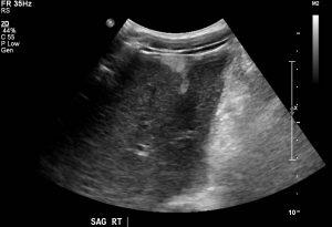
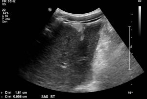
The ‘typical’ US features of hepatic hemangiomas are their small size, uniform hyperechogenicity, well-defined margin, and posterior echo enhancement. In addition, follow-up scanning only rarely shows a change in size, appearance, or detectability.
What is the reason for a hemangioma in the liver to appear echogenic?
Fat in lesion Multiple interfaces Calcification
Multiple interfaces
Their distinctive appearances at US are considered to be due to histological characteristics. They are usually composed of large blood-filled cavernous spaces, lined by a single layer of flat endothelial cells and separated by fibrous septa; multiple interfaces between the walls of the sinuses and the blood within them account for the typical hyperechogenicity seen at US.