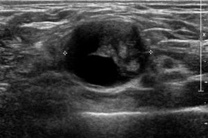
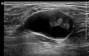
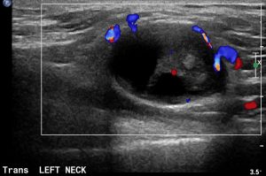
Schwannoma is a commonly encountered benign soft tissue tumor, most frequently occurring between the age of 20 and 50 years. Sites of involvement include nerves of the head and neck, the flexor surface of the upper limbs and the posterior area of the lower limbs. Sonograms of schwannoma typically show a well-defined, ovoid or round, hypoechoic, generally homogeneous solid mass with a moderate to marked posterior acoustic enhancement and eccentric positioning of the nerve trunk relative to the schwannoma. The homogeneous and decreased echogenicity of schwannomas may be explained by the uniform cellular pattern, allowing a moderate to marked sound-through transmission. A heterogeneous pattern of the mass (as observed in our case) is probably due to internal cystic or necrotic changes. The associated nerve may show thickening and loss of anisotropy. Color Doppler examination shows various degrees of vascularity ranging from minimal to abundant.