UW MEDICINE ULTRASOUND
Liver Living Donor

LIVING LIVER DONOR PROTOCOL
(UABDD)
Patient Prep: NPO for 4-6 hours.
Diagram:
Protocol:
Do the ABDOMEN DOPPLER PROTOCOL with additional documentation listed below.
Additional documentation specific for living donor ultrasound exams:
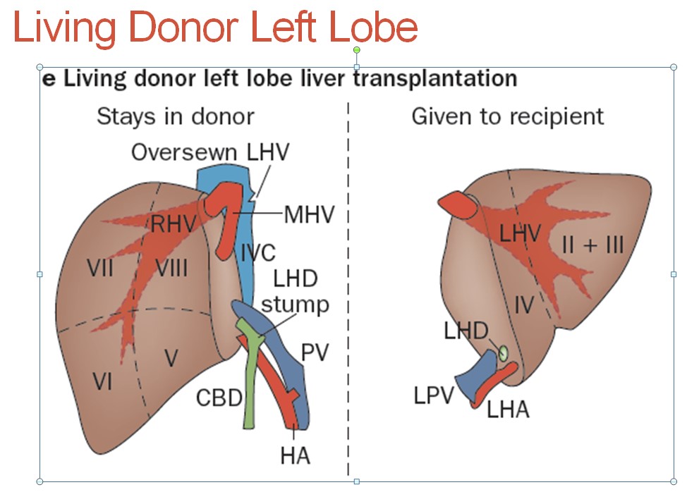
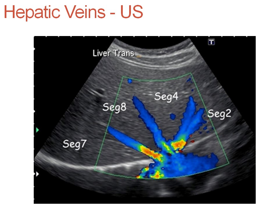
Left Hepatic Vein Documentation (subcostal view)
- Document origin of the middle and left hepatic vein in XS (2D and color Doppler).
- Measure the Left hepatic vein (inner to inner just like a yolk sac) just before it enters the IVC
- Cine-clip documentation (slow sweep please) in B-mode and color Doppler XS from superior to inferior (ROI color box should include almost the entire left lobe of the liver).
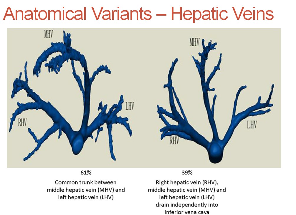
- Look for collaterals or connection between the branches of the left and middle hepatic vein – finding cross-over vein(s) that are (≥5mm) before confluence of LHV-MHV are critical for the pre-surgery work-up.
- If a collateral vein is found please document with color Doppler and 2D and measure the vein in the greatest diameter.
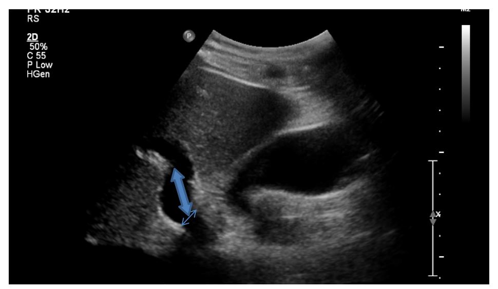
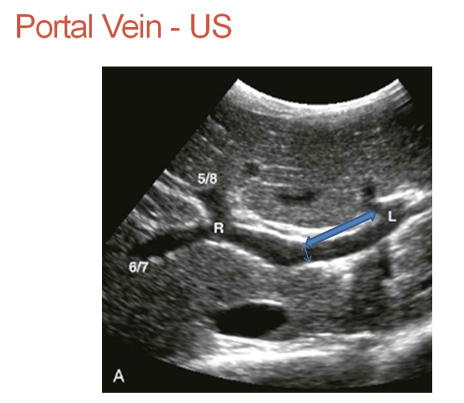
Left portal vein documentation
- Measure the diameter of the left portal vein at the origin (small arrow).
- Measure the length of the left portal vein from bifurcation until it branches (thick arrow).
Main portal vein documentation
- Measure the main portal vein diameter at the porta hepatis (small double arrow).
- Measure the length of the main portal vein up to the bifurcation (origin of the first branch) (thicker double arrow).
- 2D cine clip scanning through the main portal vein (oblique long axis view of the main portal vein) medial to the origin of the left portal vein.