
| Home |
| 3-D Echo |
| 3-D MRI |
| Applications |
| Quantitative Angiography |
| Centerline™ |
| Visual Guidance |
| Personnel |
| Publications |
| Contact Us |
Three-Dimensional Analysis of Magnetic Resonance Images
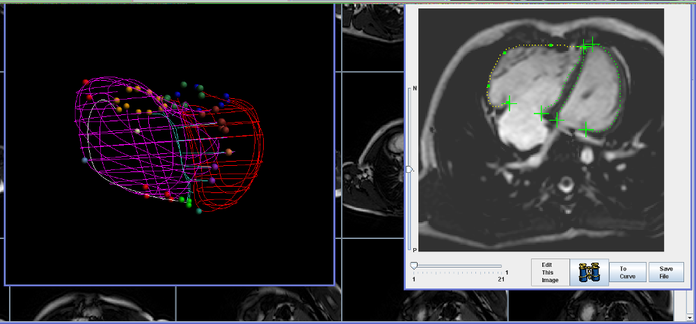
The quantitative techniques developed for three-dimensional echocardiography can also be applied to magnetic resonance images. In this study (see below), both the left ventricle (red borders) and right ventricle (pink borders) have been traced from long and short axis MRI views (seen as thumbprints in the background).
Such surface reconstructions have many potential applications. These include:
- Measurement of ventricular volume and mass
- Visualization of anatomy
- Quantification of 3D shape
- Road-mapping for procedures
- Incorporating anatomic knowledge into image processing routines
 |
|
Back to Top |
All images, text, and captions ©1998 - 2009 University of Washington, except where otherwise noted.