Anatomy & Physiology
SUMMARY: Interesting information about the sea urchin.
Sea urchins are unsegmented and penta-radial as adults. From fertilization to just before metamorphosis, the young sea urchin is bisymetrical (as are humans). At metamorphosis, the sea urchin takes on the characteristic five sided penta-radial design. (see the Development lab)
A
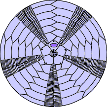
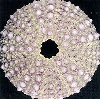
Aboral (opposite of mouth) view of a sea urchin test (shell).
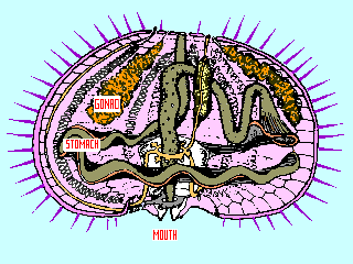
Cross sectional drawing of a living sea urchin – oral (mouth) side down.
Figure A – Aboral View
The center of the test shows the anus (very center), the 5 gonopores (5 black dots near the points of the pentagon) and madreporite (single oval in top section of pentagon). The madreporite is used to help maintain the proper pressure in the water vascular system that pressurizes the tube feet.
The tube feet pass through the pores in this section of the test.
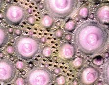
The spines are attached to the plates, between the plates where the tube feet are, on the raised "bumps" as seen below. (S. purpuratus)
images © Langstroth
Figure B – Cross-section – oral (mouth) side down
 image © Langstroth
The mouth has five teeth. The teeth and associated muscles are also called Aristotle's Lantern, after the Greek philosopher, Aristotle who first described them.
A single stomach and intestine,
and five gonads, (one shown) make up most of the bulk of the internal organs of the sea urchin. The white "squiggly" parts next to the gonad represent the water system used in the hydraulic mechanism for the tube feet (also in sets of 5). At the height of the breeding season, the gonads can represent up to 80% of the internal volume of the sea urchin.
Typically sea urchins have males and females, although occasionally it is possible to find "mistakes" that have both sexes.
Sea urchins have no "brain" as we think of it, instead they have a nerve ring near the hydraulic system used to power the tube feet.
The Tube Feet
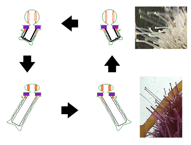
The tube feet are the primary means of locomotion for the sea urchin (along with moving the spines). Lateral movement is accomplished by means of paired muscles in the tube foot itself and extension and retraction by muscles in the hydraulic "bulb" behind each tube foot. While each "foot" may seems weak and ineffective, the large numbers of feet make up for their size.
Images redrawn from Invertebrate Zoology, by Paul Meglitsch, 1972
