
Life History
Triphasic Life History: Adapted from Images by Devon Lake |
|
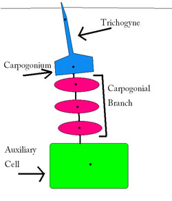
The Triphasic Life Cycle The female reproductive organ is the carpogonium. It contains two nuclei, one in the trichogyne (a narrow tube off of the main base) and then the reproductive nucleus in the base. The carpogonium is located at the end of the three-celled carpogonial branch, which is formed from inner cortical cells, which are formed by the supporting cell of the main thallus. This supporting cell also serves as the auxiliary cell once the cell is fertilized. The carpogonial branch is curved which brings the auxiliary cell towards the carpogonium, forming a procarp (Dixon, 1973) . Fertilization begins with the fusion of the spermatium to the female trichogyne. The cell walls of each of these dissolve, and the male nucleus moves down into the base of the carpogonium where it fuses with the female nucleus. Following fertilization, pit plugs form separating the carposporangial base from the trichogyne. In the base, the nucleus begins to differentiate and produces gonimoblast filaments (Kim 1976). Carposporangia develop out from the gonimoblast filaments and have diploid carpospores at their tips. This is the carposporophyte stage where an envelope of cells develops from the cortex and medulla to surround the carposporophyte which is called the pericarp. The entire structure is known as the cystocarp. The pericarp is haploid and the internal carposporophyte is diploid. These cystocarps are pushed out from the main thallus and housed in the bumpy papillae. The carpospores are then released into the water to settle and develop into the diploid tetrasporophyte, which looks the same as the gametophytes (isomorphic). The tetrasporophyte develops tetrasporangia which produce four tetraspores. Each of these is formed by a cruciate division pattern, similar to a cross (think of a circle cut into quarters and shifted apart slightly). The tetraspores are then released through holes in the cell wall (Kim 1976) and are distributed passively by water currents, settle, and grow into the gametophyte again. Lather, rinse, repeat.
References Dixon, Peter S. Biology of the Rhodophyta. Edinburgh, England: Oliver & Boyd, 1973. Kim, Dong H. A Study of the Development of Cystocarps and Tetrasporangial Sori in Gigartinaceae (Rhodophyta, Gigartinales). Germany, 1976. Lake, Devon. 1999. http://www.mbari.org/staff/conn/botany/reds/devon/home.htm Lee, Richard Edward. Phycology. Cambridge, England: Cambridge University Press, 1980. |
 Female reproductive structures