
the immortal red crust
MORPHOLOGY
|
|
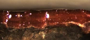
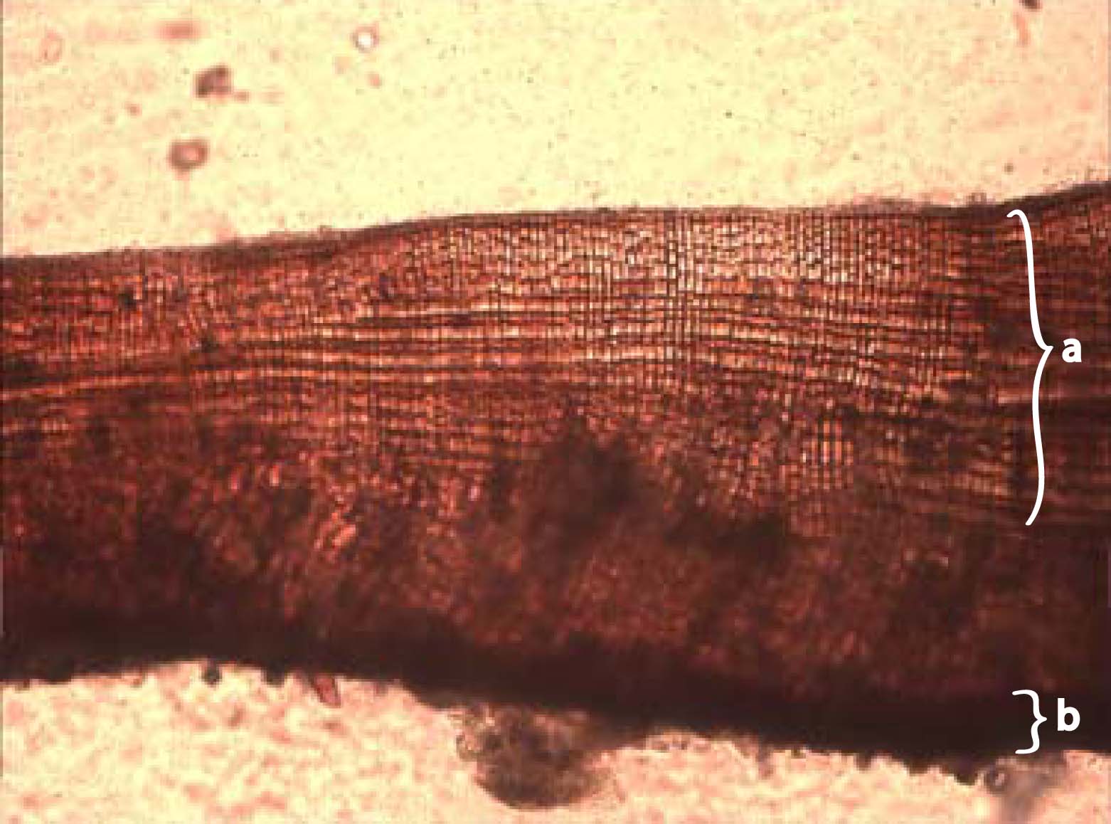
There is not a lot of diversity associated with the crustose morphology and anatomy (1). The growth of a crust is essentially 2-dimensional, but vertical growth is not an insignificant variable (see Ecology). H. rubra is one of the thinnest crusts, usually less than 150 µm thick and containing no nonphotosynthetic cells (i.e., no high-maintenance cells). In contrast, H. occidentalis can reach thicknesses of 700+ µm (2). The thallus of a crust is generally split up into the hypothallus -- the bottom layer of cells adhering to the rock -- and the perithallus -- the upper pseudoparenchymous layer of vertical filaments. Hildenbrandia spp. lack a distinct hypothallus; the entire body is pseudoparenchyma (3).
The cells of Hildenbrandia are rectangular and very small, usually 2-5 µm wide. Such a small cell size suggests a high proporition of cell wall material per unit mass and therefore a high degree of toughness (4). Another cellular feature that makes Hildenbrandia's thallus tough is the abundance of secondary pit connections among adjacent filaments (5). Unlike most pit plugs, the principal function of these connections does not appear to be the transfer of photosynthates or chemical information. The secondary pit connections in Hildenbrandia form after complete cell division (i.e., without disrupting the growth of the cell wall) and seem to be designed for increasing the coherence of the perithallus. This morphological and anatomical toughness makes it very difficult [for grazers and other disturbances] to remove Hildenbrandia from the substrate.
When Hildenbrandia is growing on softer rock, it will often incorporate little bits of the substrate up into its own thallus (6). When removing the thallus, it is not uncommon for the basal cells to bring the top layer of rock with them (see Ecology).
|
|

- A crust is a crust is a crust, but the crustose functional group demonstrates incredible physiological variation (e.g., degree of calcification). This physiological diversity is the reason why crustose algae can be found everywhere in the photic zone (see Ecology).
- Dethier, M.N. 1987. The distribution and reproductive phenology of intertidal fleshy crustose algae in Washington. Canadian Journal of Botany 65:9, pp. 1838 - 1850.
- Cabioch, J. & Giraud, G. 1982. La structure hildenbrandioïde, stratégie adaptative chez les Florideés. Phycologia 21:3, 308 - 315. Thanks to Jelte Harnmeijer (http://www.realfuture.org) for translating.
- Dethier, M.N. 1994. The ecology of intertidal algal crusts: variation within a functional group. Journal of Experimental Marine Biology and Ecology 177:1, pp. 37 - 71.
- Pueschel, C.M. 1982. Secondary pit connections in Hildenbrandia (Rhodophyta, Hildenbrandiales).European Journal of Phycology 23:1, pp. 25 - 32.
- Dethier, M.N. 2009. Personal communication.