Advanced Physical Diagnosis
Examination
  • Techniques
  • Demonstrations
Historical
Pathophysiology
Associated Evaluations
  • Patient HX
  • Physical Exam
  • Laboratory & Imaging
Differential Dx
Evidence Base
• History
• Physical Exam
• Laboratory Tests
References
Teaching Tips
[Skill Modules >> Liver & Ascites >> Differential Dx ]

Differential Diagnosis: Liver & Ascites

False Appearance

false appearance of ascites 
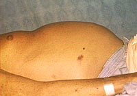
Not all patients with a protruberant abdomen or history of increasing abdominal girth have ascites.

The following conditions may give a false positive appearance of ascites:

  • The obese patient with a lax anterior abdominal wall may have a protuberant umbilicus (everted umbilicus sign)
  • Metastases to the liver - in this case the firm liver holds the abdomen outward. Gaseous distention due to bowel obstruction
  • Large pelvic mass
  • Massive hepatosplenomegaly as in lympho-proliferative conditions

Abdominal distention from the above causes can usually be distinguished from ascites by examination and percussion of the flanks. In ascites, the fluid falls into the lateral and lower peritoneal spaces when the patient is supine, giving bulging flanks and flank dullness. The absence of flank dullness on careful examination of a patient with abdominal distention excludes ascites with 90% accuracy (link to Evidence-base section)

back to top

Etiology of Ascites

Cause % of all cases
Chronic liver disease (cirrhosis or chronic hepatitis) 80%
Underlying malignancy 10%
Congestive heart failure 3-5%
Cirrhosis and another etiology 5%
Infection: Tuberculous peritonitis 1%
Infection: Chlamydia (*Fitz-Hugh-Curtis syndrome) <1%
Infection: **HIV-related ascites <1%
Renal disease (nephrotic syndrome, dialysis-related <1%
Fulminant hepatic failure <1%
Pancreatic <1%
Biliary <1%
Chylous <1%
* perihepatitis rarely due to gonococcus
**Note: ~5% of pts with AIDS have ascites detected by ultrasound; many etiologies

back to top

Ascites may be differentiated by the nature of the fluid

  • The traditional characterization of ascitic fluid as "transudative" or "exudative" is no longer used.
  • Although many laboratory chemistry tests may be performed on ascitic fluid, among the most useful is the serum - albumin gradient, calculated as follows:

    Serum albumin - ascites albumin (note: this is subtraction, not a ratio)

The utility of the albumin gradient is based on the concept of oncotic-hydrostatic balance. If the patient has a high portal pressure, there will also be a high oncotic gradient, with albumin contributing the major component of serum proteins. There are large differences between serum and ascitic fluid albumin concentrations in portal hypertension, and in > 90% of cases, will be associated with a high gradient.

Etiology based upon serum - ascites albumin gradient

Although many laboratory chemistry tests may be performed, among the most useful is the serum - albumin gradient, as follows:

Serum albumin - ascites albumin

Etiology based upon serum - ascites albumin gradient
High albumin gradient
(>1.1 g/dl)
Low albumin gradient
(<1.1 g/dl)
Cirrhosis Peritoneal carcinomatosis
Alcoholic hepatitis Tuberculosis
Cardiac ascites Tuberculosis
Massive liver metastases Biliary ascites without cirrhosis
Fulminant hepatic failure Nephrotic syndrome
Hepatic vein thrombosis (Budd-Chiari syndrome) Ascites associated with connective tissue disease
Portal vein thrombosis Ascites associated with bowel ischemia/infarction
Veno-occlusive disease  
Acute fatty liver of pregnancy  
Myxedema  
"Mixed ascites"  

For more discussion on pathogenesis of ascites, check the references.

back to top