“…very small living creatures in rain water.”
The evolution course I took as an undergraduate was co-taught by two professors, one who studied butterflies and the other (my advisor at the time) who studied viral ecology and evolution. For one of the classes, the butterfly professor brought in models and pinned specimens to show off their beautiful patterns. My advisor decided he couldn’t be one-upped. “I had organism envy,” he explained to us as he passed around Petri dishes on which some viruses had been grown.
The organisms we study are all interesting and dynamic, but that can be easy to forget when they’re things that are not publicly “charismatic”, not bright and colorful or cute and cuddly, or just too small to see. For example, I only see bacteria when there are millions of them in one place, enough to turn liquid turbid or form a small, moist-looking dab on a Petri dish. At this scale, it can seem as though things are fairly static. The liquid only becomes so turbid. The moist dab only grows to a certain circumference.
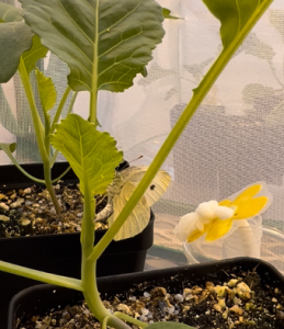
But during a synthetic biology course from Eric Klavins (UW Electrical Engineering), I had a chance to see my bacteria as individuals, rather than a collective. We decided that we wanted to characterize how individual bacterial cells were growing, so we turned to microscopy. Looking through the microscope, where little oblong cells zipped about or turned abstractedly in circles, was like discovering an entire new world. Oh—this is what my organism looks like! This is how it behaves! Then we added antibiotics and watched the cells explode, which was way more fun than it should have been.
If you cannot see the video, click here to view a larger version.
Video credit: Sonia Singhal, Rashmi Ravichandran, Gregory Rowe, Rob Egbert
Time series taken over 16 hours, with one hour of growth preceding the addition of antibiotic (ampicillin). Original (and larger) video here.