|
Image #2 (#2of 14 found) |
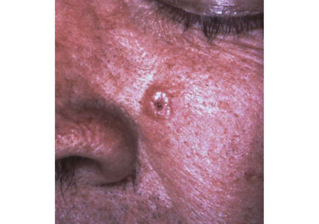
| Basal cell carcinoma: Nodular type |
Detail:
Clinical: dome-shaped, pearly or flesh-colored papule or nodule with telangiectasia; center may be ulcerated. Typically occur in sun exposed areas of fair skinned individuals (also can occur in Hispanics). They are slow growing but can outgrow their blood supply leading to ulceration and bleeding. Rarely metastasize.
Pathology: Aggregates of basaloid cells often contiguous with the epidermis. Neoplastic cells are uniform in size and possess large nuclei and scant cytoplasm. Cells in the periphery of the tumor islands align in a parallel array that has been termed palisading. A characteristic cleft is often present between neoplastic cells and fibrous stroma.
|
|
| S Taylor © UT Southwestern |
|