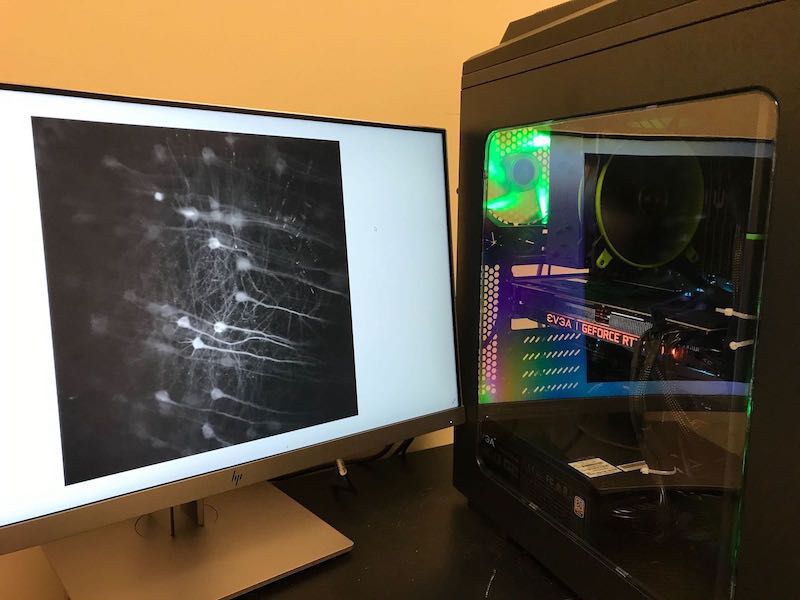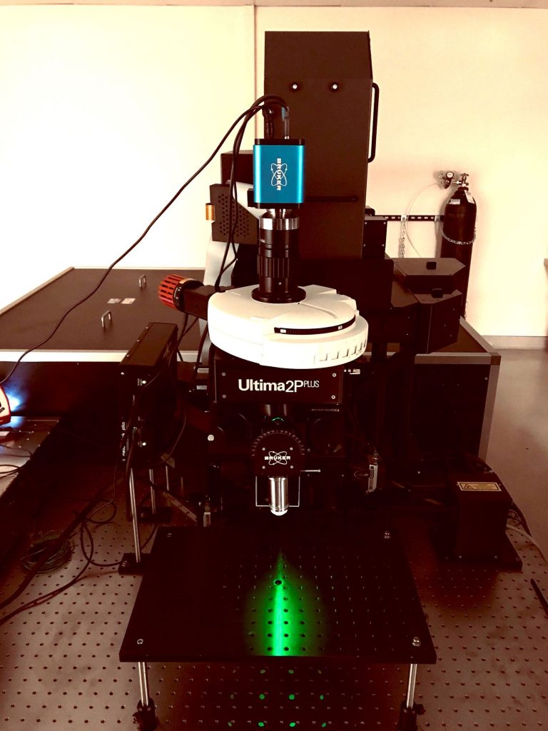
Bruker Ultima 2p Plus Microscope
This new two-photon microscope addition to the imaging core extends the users’ ability to perform high-resolution single cell-resolution functional imaging coupled with holographic optogenetic stimulation. This state-of-the-art microscope features high customizability and is fit with a spatial light modulator (SLM) optical component that allows for holographic photostimulation across 100’s of user defined regions of interest (ROIs) distributed across the x, y, and z axes. This microscope is excellent for experiments requiring stimulation of specific spatially segregated neurons to understand the underpinnings of behavior and brain dynamics.
Olympus FVMPE-RS Multiphoton Microscope
The core houses Olympus two-photon microscopes that feature high-sensitivity, high-resolution calcium imaging capabilities with a user-friendly, well-supported acquisition program. Coupled with the MaiTai-DeepSee Laser, this microscope is suitable for a broad range of imaging experiments using common fluorophore colors from yellow to red. These microscopes are ideal for in-vivo behavioral experiments with single-cell resolution imaging.
Spectra-Physics Spirit HE Laser
This laser is used with the Bruker Ultima 2p Plus microscope for two-photon
holographic stimulation. This fixed wavelength (1040 nm) laser has ultra-high
high power (8 W) to delivery ultra-short high energy pulses needed for
two-photon photo stimulation. The high-power output of this laser allows for
two-photon stimulation across 100’s of user defined regions of interest (ROIs).
Spectra-Physics Insight X3 Ti:Sapphire Laser
This laser features a broad 680 nm to 1300 nm continuous, gap free tuning from a single source, and is ideal for in vivo two photon imaging with the Bruker 2p Plus microscope. The InSight X3 delivers high average and peak power levels across the tuning range, including critical near infrared wavelengths above 900 nm for deepest penetration in-vivo tissue for use with GCaMP and next generation red-shifted calcium indictors.
Spectra-Physics MaiTai-DeepSee Laser
This laser powers the Olympus FVMPE microscopes and features a wide tuning range for efficient excitation of common fluorophores. This laser delivers excellent levels of peak power and allows for adjustable dispersion compensation to accommodate imaging at different wavelengths.
Tucker-Davis Technology RZ5P Fiber Photometry Rig
This recording system allows for simultaneous acquisition of multi-channel, multi-region brain recordings in freely-moving, behaving animals. The rig is highly mobile and is able to integrate with a diverse set of behavioral experimental paradigms. This set up is ideal for experiments focused on region-level recordings in freely-behaving animals.
Olympus FV3000 Confocal Microscope
This microscope features the latest technologies for fluorescence confocal imaging and streamlined acquisition software through the Olympus Fluoview software (same as what is used with the FVMPE-RS multiphoton microscope. This microscope is ideal for high-resolution imaging of fixed tissue on slides.
Zeiss ApoTome.2 Widefield Microscope
This microscope also serves to imaging fluorescent slides and also has the added capability for structured illumination of thick samples. As a widefield microscope, large areas of the sample can be imaged in quick succession for fast acquisition and processing of stitched composite images.
Analytical and Computational Resources
You collected beautiful videos of hundreds of neurons, now what? Please click the following link to learn about analysis resources for preprocessing the data, extracting activity from the neurons, and linking them to behavior and your functional calcium imaging experimental paradigms:
https://github.com/zhounapeuw/NAPE_imaging_analysis

Dr. Charles Zhou
Director
Please reach out for training and consultations for core equipment usage.