In the U.S., birth defects are the leading cause of death in children (National Vital Statistics Report 2004). A surprising 3% of all newborns exhibit major malformations; of these, one fifth clearly result from genetic disorders. Our goal is to define the mechanisms and molecules that control these developmental processes, that we can develop diagnostic tools for identifying risks factors and, in the long run, evaluate the efficacy of potential treatments.
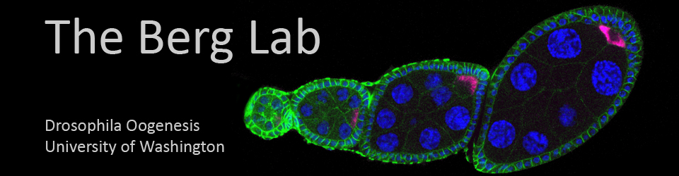
What are the mechanisms and molecules that control development? Importantly, genetic and genomic studies reveal that developmental processes are similar in all animals. Thus, we use a simple Drosophila model to investigate how flat epithelial sheets are patterned into distinct cell types and how these cell types change shape and rearrange to make a pair of simple tubes called dorsal appendages (Dorman et al. 2004; reviewed by Berg 2005). This process occurs in the follicle cell layer that surrounds the oocyte and is tied to establishing dorsal/ventral polarity in the embryo. In vertebrates, tube formation produces the heart, kidneys, lungs, gut, and neural tube; dorsal appendage formation resembles these more complex processes but is easier to study because it does not involve cell division or cell death. We also have sophisticated genetic tools for manipulating gene function, a culture system for imaging events live, and a battery of markers for following the fate and behavior of the cells.
We are interested in six broad questions:
How do genes control the differences in size and shape of the tubes?
Genetic variants exhibit striking differences in morphology that mimic natural shapes throughout the animal kingdom. Some variations affect patterning and alter the number of cells fated to produce the tubes; others affect morphogenesis and alter the cell biological processes that produce the tubes (Berg 2005). What are these genes and how similar are their roles in different developmental processes?
How does a gradient of signaling information resolve into a sharp boundary between two distinct cell types?
Several signaling pathways contribute to defining the two types of cells that make the dorsal appendage tubes. EGF and BMP signals are expressed in a graded fashion in the dorsal anterior follicle cells. These molecules induce Notch and Wingless signaling, which subdivide the primordium and establish a boundary between the two cell types (Ward and Berg 2005; Ward et al. 2006). How do these pathways refine the pattern within the epithelium?
How do the two cell types coordinate their efforts to produce a tube?
Cells expressing high levels of the transcription factor Broad constrict their apices and undergo convergent extension to form the roof and sides of the tube. Cells expressing the protease Rhomboid elongate dramatically and zipper together to seal off the floor of the tube (Dorman et al. 2004). Subsequent shape changes and rearrangements produce the final form of the tube. How does each group of cells coordinate their activities, within each group and between the two cell types?
What other genes contribute to tube formation?
Although we know dozens of genes that affect the patterning and morphogenesis of the dorsal-appendage tubes, we have only just begun to understand this complex process. We are using genetic, genomic, and proteomic approaches to identify new genes that regulate development.
How do different species adapt their patterning and morphogenesis processes to generate dorsal appendage tubes appropriate for their life histories?
Our studies thus far reveal remarkable similarities with mechanisms in D. melanogaster as well as striking differences in more distant species.
How do these differences evolve?
Drosophila provides an outstanding system for investigating the fundamental processes that regulate development and for identifying the genes that, when mutated, contribute to birth defects.