|
|
|
Image Acquisition
Rapid Free Scanning for Multiplane Acquisition
3D Tracking and the Imaging System
Rapid Free Scanning for Multiplane Acquisition
Imaging is performed using a phased-array scan head and a commercial ultrasound machine. The 3D image set is acquired over five or six 6-10 sec periods of held end-expiration. The duration and number of scans is adjusted according to each patient's breath-holding ability and image quality. The patient is instructed not to move during imaging or between scans, and to suspend respiration at the same level of expiration during each scan. Each image set can thus be acquired in as brief a time as a single heartbeat14.
The first scan starts with the parasternal long axis view; the probe is then rotated to short axis and the probe is angulated from the base to mid LV (see Figure 1). The second and 3rd through 5th scans are performed as depicted in Figures 2 (short axis) and 3 (apical). At the apical window, three separate scans are used to cover 4-chamber/5-chamber, long-axis, and 2-chamber view sets, respectively. The sixth scan is optional, and can supplement any view requiring additional coverage.
Additional scans may be performed for specific research interests, such as imaging the right heart. Each scan may be reviewed after acquisition and either saved or repeated.
 |
|
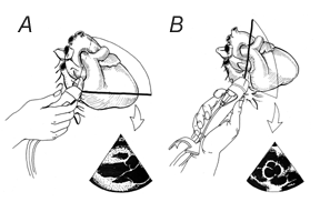
A: Starting orientation for rapid free scanning in the parasternal position. View is parasternal long axis. B: Ending position for rapid free scanning in the parasternal position. Probe is rotated from the starting position 90 degrees clockwise from the operator's viewpoint, to display parasternal short axis at the aortic valve level. |
 |
|
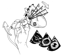
B: Rapid free scanning for parasternal short axis coverage. Initial view is basal short axis at aortic valve level. Probe is tilted cephalad, aiming field down towards apex. Ending position varies, depending on heart size, but usually mid-ventricle. For apicalmost views, tilting scan procedure may be repeated from point B. |
 |
|
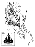
C: Rapid free scanning in the apical position. Initial view is apical 4-chamber. Probe is rotated clockwise for 5-chamber view and back again to 4-chamber starting point (scan 3). For 2-chamber views, probe is rotated counterclockwise and field is tilted inferiorly (scan 4); for long-axis, tilt is cephalad (scan 5). |
3D Tracking and the Imaging System
A magnetic field system (Flock of Birds® model 6D-FOB, Ascension Technology Corp., Burlington, VT) is used to track the ultrasound scanhead. The system consists of a magnetic field transmitter, a receiver, and electronic circuitry which interfaces with a computer (see below). The transmitter sequentially generates three orthogonal magnetic fields which are sensed by the receiver and used to compute the receiver's position (x, y, z) and orientation (azimuth, elevation, roll) in space with respect to the origin of the transmitter. During imaging, the transmitter is maintained in a constant position, and the receiver is attached to the ultrasound scan head. Images are digitized directly using a framegrabber (Mutech, Billerica, MA) under the control of a personal computer (Pentium®) running custom software. Images are registered with position data, hemodynamic parameters, and scanning parameters such as image depth.
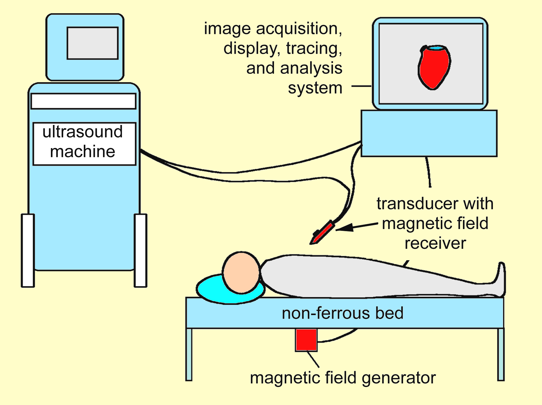 |
| Schematic of the 3D Echo laboratory setup. The magnetic locator device components are adapted to fit onto standard ultrasound imaging equipment. Simultaneous ECG recordings (not shown) are recorded, transferred, and indexed with the appropriate image frames. |
Back to Top
|