
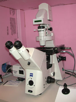
Fluoview-1000 Confocal Microscope (Olympus)
The FV-1000 is a laser scanning confocal microscope built on an IX-81 inverted microscope (Olympus) equipped with a motorized stage, epi-fluorescence and differential interference contrast (DIC). The FV-1000 has 6 laser lines with 4 detectors for fluorescence plus a transmitted light detector. All 5 detectors may be acquired with simultaneous or sequential scanning. It is possible to collect up to 6 channels of fluorescence by automated dichroic switching.
| Objective | NA | Working Distance | Immersion | Purpose |
|---|---|---|---|---|
| 4X UPlanSApo | 0.16 | 13 mm | Air | Orientation, overview |
| 10X UPlanSApo | 0.40 | 3.1 mm | Air | Orientation,low mag. |
| 20X LWD1,3 | 0.45 | 7.8 mm | Air | Culture plates |
| 20X UPlanSApo | 0.75 | 600 µm | Air | Medium resolution |
| 40X UPlanFL N | 1.30 | 200 µm | Oil | High resolution |
| 60X PlanSApo | 1.35 | 150 µm | Oil | High resolution |
| 100X UPlanSApo | 1.40 | 130 µm | Oil | High resolution |
| 60X UPlanSApo2,3 | 1.20 | 280 µm | Water | Live imaging |
FV-1000 laser lines:

Marianas Microscope
This imaging system was integrated by Intelligent Imaging Innovations, Inc. (“3I", Denver, CO) and controlled by their Slidebook software. The Marianas is designed to provide a stable, highly automated platform on an inverted microscope for critical imaging applications such as timelapse, deconvolution, FRET and ratiometric imaging as well as high resolution brightfield and fluorescence image capture.
| Objective | NA | Working Distance | Immersion | Pixel Size1 µm/pixel | Purpose |
|---|---|---|---|---|---|
| 5X Plan NeoFluor | 0.15 | 13.6 mm | Air | 1.249 | Orientation |
| 10X Plan NeoFluor | 0.30 | 5.6 mm | Air | 0.624 | Low mag. imaging |
| 20X Plan Apochromat | 0.75 | 610 µm | Air | 0.314 | Medium resolution |
| 25X LCI-PlanNeoFluor2,4 | 0.80 | 600 µm | Multiple | 0.244 | Specialty |
| 40X Plan NeoFluor | 0.75 | 500 µm | Air | 0.156 | Medium resolution |
| 63XW3 C-Apochromat3,4 | 1.20 | 240 µm | Water | 0.099 | Live imaging |
| 63X Plan Apochromat | 1.40 | 180 µm | Oil | 0.099 | High Resolution |
| 100X Plan Apochromat | 1.40 | 170 µm | Oil | 0.062 | High Resolution |
Notes:
Nikon Optiphot
A basic, manual upright microscope capable of brightfield, phase contrast and epi-fluorescence. The CCD camera uses a tunable filter to collect full resolution images of fluorescent labels as well as in RGB color for non-fluorescent specimens. It is popular for collecting RGB images of conventional brightfield stains such as H&E, crystal violet or DAB. It is often used to inspect specimen label quality before attempting to image on the advanced instruments.
| Objective | NA | Working Distance | Immersion | Pixel Size µm/pixel | Pixel Size1 pixel/µm |
|---|---|---|---|---|---|
| 1X Plan | 0.04 | 13.6 mm | Air | 8.40 | 0.12 |
| 2X Plan | 0.05 | 5.6 mm | Air | 4.24 | 0.24 |
| 4X EPlan | 0.10 | 610 µm | Air | 2.12 | 0.57 |
| 10X EPlan | 0.40 | 600 µm | Air | 0.87 | 1.15 |
| 20X EPlan | 0.04 | 500 µm | Air | 0.43 | 2.31 |
| 40X EPlan | 0.65 | 240 µm | Air | 0.21 | 4.78 |
| 40X Fluor2,4 | 0.85 | 180 µm | Air | 0.21 | 4.78 |
| 100X3,4 | 0.90-1.25 | 170 µm | Oil | 0.09 | 11.55 |
| 100X Fluor2,4 | 1.30 | 170 µm | Oil | 0.09 | 11.55 |