| Research | |||||||||||||||||||||||
|
Milestones in Mass Spectrometry |
|||||||||||||||||||||||
|
When an architect presents house blueprints to a construction crew, the house is far from being considered complete and habitable. Construction workers build the house according to the set of blueprints, and then the plumbers and interior decorators finish the job.
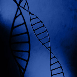
Walsh, professor emeritus of biochemistry, joined the UW faculty in 1959 after receiving his Ph.D. in biochemistry from the University of Toronto. He was chair of the Department of Biochemistry from 1992 to 2000, when he retired In September 2002 the International Association for Protein Structure Analysis and Proteomics presented the Pehr Edman Award to Walsh for his contributions to protein chemistry, protein structure analysis, and proteomics. "Proteins are the working machines of the cell," said Walsh. "Heart cells display different proteins from brain cells, and so the cells differ in function. The challenge is to identify which proteins actually occur inside each kind of cell. With 30,000 possibilities, fast methods are necessary." By 1980 DNA sequencing was replacing the Edman degradation, the classic, although laborious, way to determine amino acid sequences of proteins by taking them apart one amino acid at a time. "I did Edman degradations for 30 years," said Walsh. "When DNA sequencing came on the scene, we at the UW began to focus on other ways to characterize proteins, and the mass spectrometer became the tool of choice."
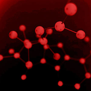
"It's one thing to translate the genetic information," said Walsh "It's quite another to assemble mature, functioning proteins. Back to the house analogy - a construction company can build a house from blueprints, but the house is not a functioning residence until the company put locks on the doors and toilets on the floors. We focused on the chemical changes in the proteins that occur after they are translated from the DNA, the so-called post-translational modifications." The mass spectrometer became the ideal analytical tool, Walsh said, because all post-translational modifications either add mass, like a phosphate group, or they take away mass, like a segment of the protein. For instance, proinsulin only becomes insulin when an enzyme breaks some molecular bonds and removes a segment, thereby decreasing its mass. From a change in the mass of a protein, or a fragment of it, the nature of the attached group or removed segment can be deduced, and much can be learned of the function or control of the protein. "With a mass spectrometer you can actually see the change in molecular mass," said Walsh. "For instance, Krebs and Fischer's Nobel-Prize winning discovery of phosphorylation in the 1950s would have been easier by mass spectrometry. When a phosphate group attaches to a protein an 80-dalton increase in the mass of a segment of a protein signals that it has been phosphorylated." Walsh's lab at UW closed in 2000. His mass spectrometers are still available in the School of Pharmacy and the Department of Chemistry. |
|||||||||||||||||||||||
|
© 2003 - 2004 UW Medicine
Maintained by UW Health Sciences and Medical Affairs News and Community Relations Send questions and comments to drrpt@u.washington.edu |
|||||||||||||||||||||||