[Skill Modules
>>
Liver & Ascites
>>
Physical Exam
]
Physical Exam: Liver & Ascites
There are many other physical findings to look for in the patient with ascites:
Skin:
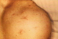
- Palmar erythema
- Spider angiomata - most commonly on the trunk and upper extremities
- Caput medusae (dilated venous pattern over the right upper abdomen)
Fluid Overload:
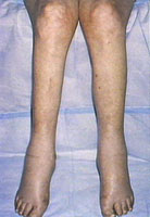
- Peripheral edema
Note: edema in ascites due to liver or heart disease is usually confined to the lower extremities only; if present also in upper extremities and hands, consider renal disease and nephrotic syndrome.
- Jugular venous distension (see JVP below)
- Pulmonary crackles (suspect CHF)
- Cardiac S3
- Positive hepatojugular reflux (link to neck veins module associated examination segment for description of exam maneuver)
Jugular venous pressure (JVP)
- When elevated, suspect right-sided heart failure
- If high JVP, also examine jugular waveforms further for constrictive pericarditis and check pulsus paradox
Lymphadenopathy; other signs of malignancy
Consider signs of other uncommon etiologies of ascites:
- Hypothyroidism
- Thyroid may be enlarged; skin dry with brittle hair; tongue enlarged (macroglossia), peri-orbital edema, delayed peripheral deep tendon reflexes (delay most prominent in return phase of reflex exam)
- Hemochromatosis
- Skin grayish or bronze and appears dirty
- Degenerative arthritis of extremities (usually hands and fingers, especially affected are PIPs of the middle and ring fingers)
- Wilson's disease (generally always presents before age 50)
- Eye: Kayser-Fleisher ring: brownish-green ring near limbus edge of iris - represents copper deposition in Descemet's membrane (has high sensitivity and specificity, although may need slit lamp to see)
- Funduscopic: "hyaloid" or colloid bodies - getanitous appearance on edge of disc that obscures the disc border (mimics papilledema); can give pt visual filed defects
- Nails: bluish discoloration of the lunula (termed azure lunule; not specific)
|