<--Previous Up Next-->
P75cochlea3D
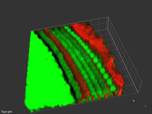
This volumetric projection of the apical turn of a P5 mouse cochlea labeled for p75 (red) and nuclei (green). The characteristic arch formed by the union of the inner (left) and outer (right) pillar cells (red) is seen to the left. The large green nuclei to the left of the pillars are of inner hair cells. The red nerve fibers may be seen below the outer hair cells (OHC), partly obscured by the the OHC nuclei, and extending upwards around the OHC. The Claudius cells at far right are intensely labeled for p75. Dr. Elizabeth Oesterle.