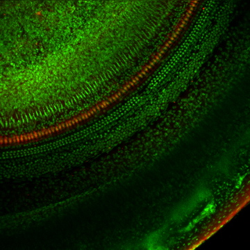
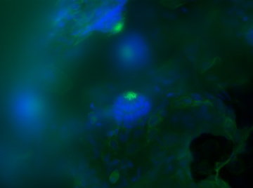
 03-6403T1 10xBP First turn of a mouse cochlea labeled to show all cell nuclei (green) and calretinin (red) which occurs in neurons and in Inner Hair Cells (IHC). The single row of Inner Hair Cells arcs across the middle of the image. The 3 rows of Outer Hair Cell nuclei are observed in their characteristic arrangement paralleling the arc of IHC. Whole mount label embedded in epoxy resin, imaged with Bio-Rad MRC-1024 confocal microscope, 10x objective (Nikon). Glen MacDonald. |
 311T2MLEproj High resolution imaging of 3 inner hair cells (IHC) innervated by dendrites of the neurons in the spiral ganglion. Calretinin (red) labels the IHC while nerve fibers were labeled for neurofilaments (green). Afferent fibers may be seen extending to the left of the IHC, en route to the outer hair cells and support cells. Nuclei appear blue. Whole mount label embedded in epoxy resin, imaged with widefield microscope, 63x objective (Zeiss) and deconvolved (Huygens software). Glen MacDonald. |
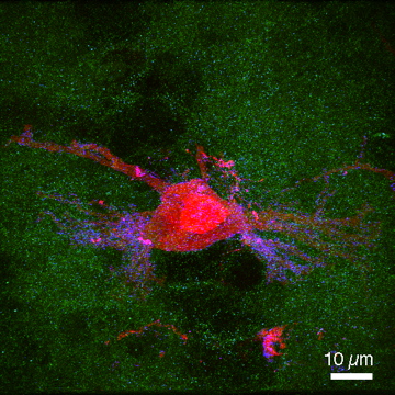 NL_Synaptophysin Single neuron in avian N. lamaris filled with rhodamine-conjugated dextran (red) surrounded by fluorescent label for the pre-synaptic protein, synaptophysin (green). The blue dots represent putative colocalization of synaptophysin along the dendrites. 100X/1.4 (Nikon) on a Bio-Rad MRC-1024, deconvolved by Huygens, masking and projection by Slidebook. Staci Sorensen and Rachael Nehmer. |
 NM phalloidin TOPRO PIC This is a picture of zebrafish neuromasts stained with flourescent dyes, Phalloidin (green) and Topro-3 (blue). Keri O'Connell. |
 mtgmtrtp3BP adj Raw images of live neuromast hair cells from a zebrafish. Cells appear outlined in yellow as mitochondria are labeled for a fluorescent indicators of all mitochondria (green) and mitochondria with a high electropotential gradient (red). The raw images appear blurred from out of focus light and optical effects of the sample. Some mitochondria appear of one color due to organelle motion during the image acquisition. Brightest point projection of 72 images collected by widefield fluorescence with a 63X water immersion objective (Zeiss). Glen MacDonald. |
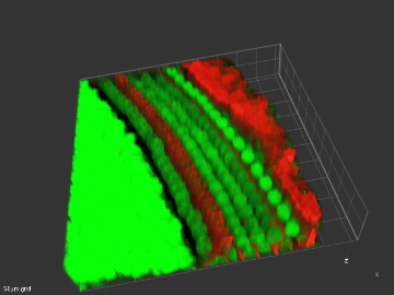
 MTRMTGTP3ci bp The same optical volume displayed above, but processed by deconvolution using Slidebook. Removal of blur restores the resolution of the image. Mitochondria are distinctly visible. Glen MacDonald. |
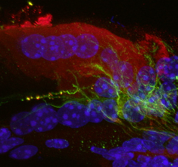
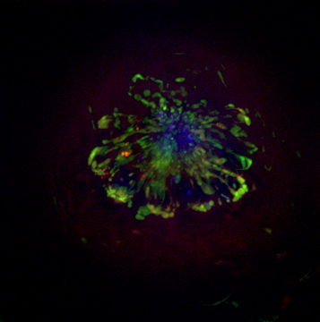
 P75cochlea3D This volumetric projection of the apical turn of a P5 mouse cochlea labeled for p75 (red) and nuclei (green). The characteristic arch formed by the union of the inner (left) and outer (right) pillar cells (red) is seen to the left. The large green nuclei to the left of the pillars are of inner hair cells. The red nerve fibers may be seen below the outer hair cells (OHC), partly obscured by the the OHC nuclei, and extending upwards around the OHC. The Claudius cells at far right are intensely labeled for p75. Dr. Elizabeth Oesterle. |
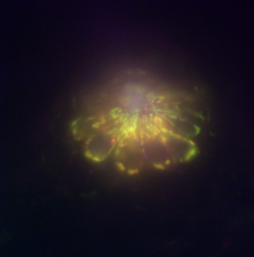
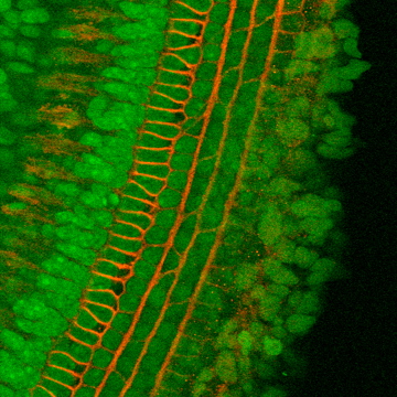 P75cochleaBP A brightest point projection from a confocal image volume of the apical turn of a mouse cochlea, aged P5. This shows the Organ of Corti stained for p75 receptors (red) and nuclei (green). The U-shaped structures are the feet of inner and outer pillar cells, while cicumferential nerve fibers run between the 3 rows of outer hair cells. Dr. Elizabeth Oesterle. |
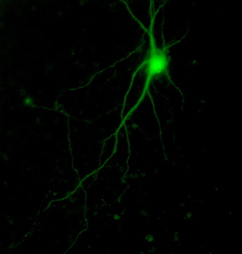 05-6809_6p_fitc GFP-expressing auditory brainstem cell of a five day old animal after 7 days in culture. The GFP plasmid was electroporated after 3 days in vitro. |
Photo Gallery
- Home
- Lab Information
- Lab Safety
- Calendar
- Personnel
- Fish Hair Cell Group
- Contact Us
- Research Overview
- Photo Gallery
- PBio Seminars
- Links
- Protocols
- Occupational Health
- Digital Microscopy Center (DMC)
Equipment reservations
PURCHASING FORM
Archives