<--Previous Up Next-->
P75cochleaBP
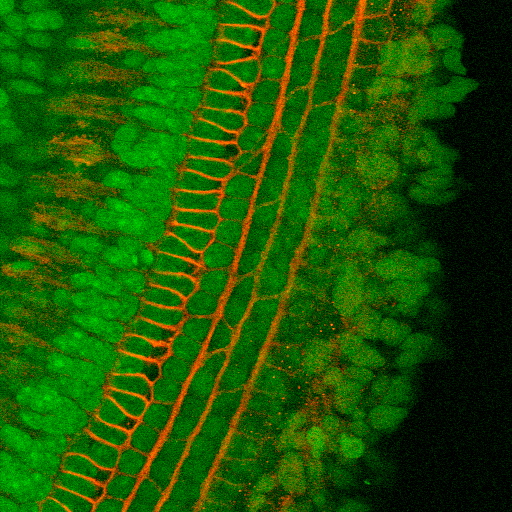
A brightest point projection from a confocal image volume of the apical turn of a mouse cochlea, aged P5. This shows the Organ of Corti stained for p75 receptors (red) and nuclei (green). The U-shaped structures are the feet of inner and outer pillar cells, while cicumferential nerve fibers run between the 3 rows of outer hair cells. Dr. Elizabeth Oesterle.