
Image1
Image2
Image3
Image4
Image5
Image6
Image7
Image8
Image9
Image10
Image11
Image12
Image13
Image14
Image15
Image16
Image17
Image18
Image19
Image20
Image21
Image22


Horizontal cells of the zebrafish retina.
Image by Leanne Godinho and Philip Williams.
Cyan: Bodipy staining of cell nuclei. Yellow: Cx55.5:M-YFP horizontal cell precursors. Red: Outer plexiform layer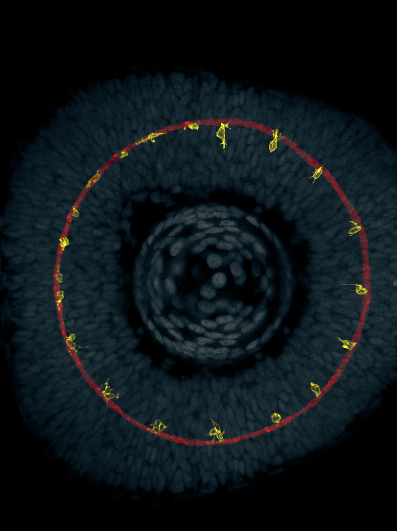
Image by Leanne Godinho and Philip Williams.
Cyan: Bodipy staining of cell nuclei. Yellow: Cx55.5:M-YFP horizontal cell precursors. Red: Outer plexiform layer
Zebrafish embryo.
Image by Philip Williams.
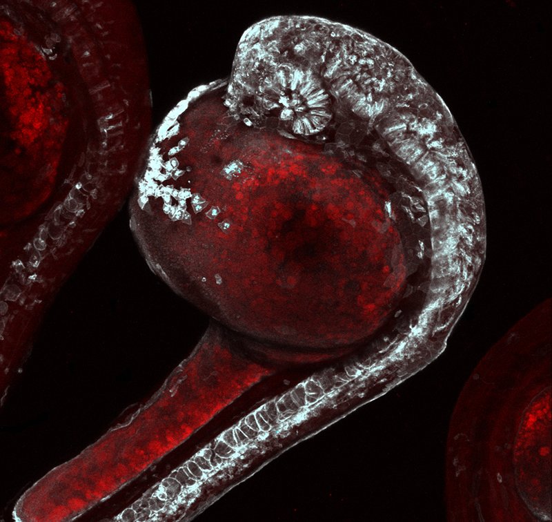
Image by Philip Williams.
Cross section of a zebrafish retina.
Image by Philip Williams.
From a 5-day old zebrafish expressing fluorescent protein and immunostained for major retinal cell types.
Image by Philip Williams.
From a 5-day old zebrafish expressing fluorescent protein and immunostained for major retinal cell types.
Zebrafish eye.
Image by Philip Williams.
Purple: Cone photoreceptors. Orange: Mueller glia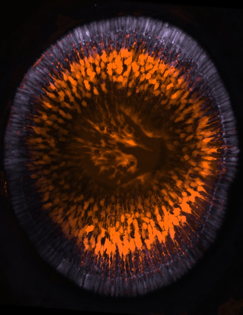
Image by Philip Williams.
Purple: Cone photoreceptors. Orange: Mueller glia
Multi-ribbon synapse in tetanus toxin-expressing retina.
Image by Rachel Wong, Ed Parker and Daniel Kerschensteiner.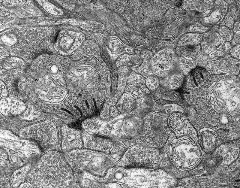
Image by Rachel Wong, Ed Parker and Daniel Kerschensteiner.
Horizontal cell contacting photoreceptors in the mouse retina.
Image by Timm Schubert.
Yellow: Horizontal cell filled intracellularly with fluorescent dye. Purple: Cone photoreceptor axon terminals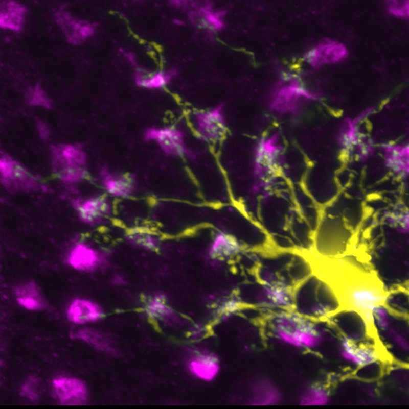
Image by Timm Schubert.
Yellow: Horizontal cell filled intracellularly with fluorescent dye. Purple: Cone photoreceptor axon terminals
Synaptic contacts between bipolar and ganglion cells in the mouse retina.
Image by Josh Morgan.
Red: Axon terminal of a cone bipolar cell. Cyan: Ganglion cell dendrites. Yellow: Postsynaptic density protein PSD95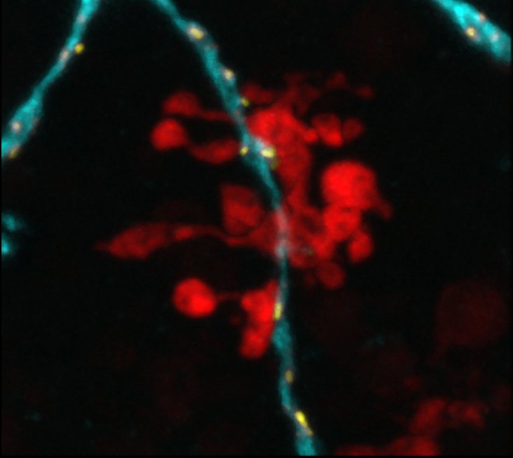
Image by Josh Morgan.
Red: Axon terminal of a cone bipolar cell. Cyan: Ganglion cell dendrites. Yellow: Postsynaptic density protein PSD95
Neighboring ganglion cells labeled biolistically in the mouse retina.
Image by Luca Della Santina.
Cyan, Yellow: Pseudocolored A-type ON ganglion cells expressing a cytosolic fluorescent protein.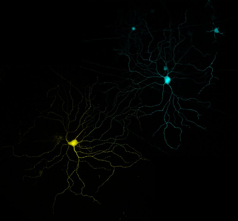
Image by Luca Della Santina.
Cyan, Yellow: Pseudocolored A-type ON ganglion cells expressing a cytosolic fluorescent protein.
Zebrafish "has" mutant eye.
Image by Jung-Hwan Choi.
Red: cellular membranes. Yellow: GFP-expressing ganglion cells.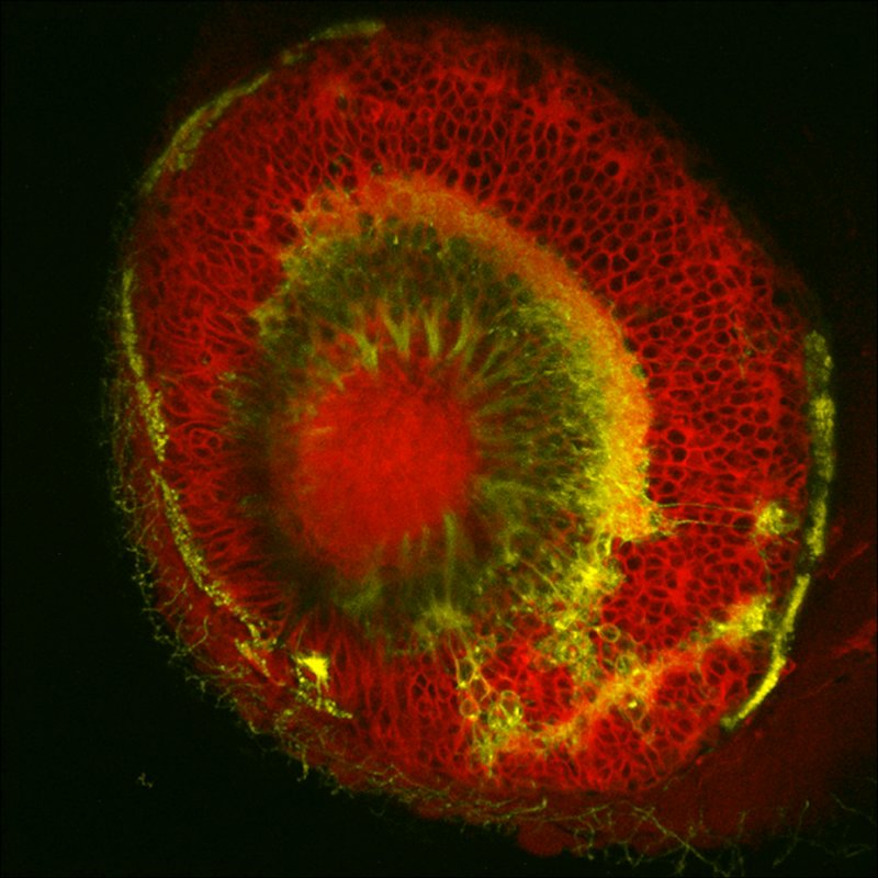
Image by Jung-Hwan Choi.
Red: cellular membranes. Yellow: GFP-expressing ganglion cells.
Retinal ganglion cell contacting ON bipolar cells.
Image by Josh Morgan.
Red: ON bipolar cells terminals. Cyan: Ganglion cell dendrites. Yellow: Postsynaptic density protein PSD95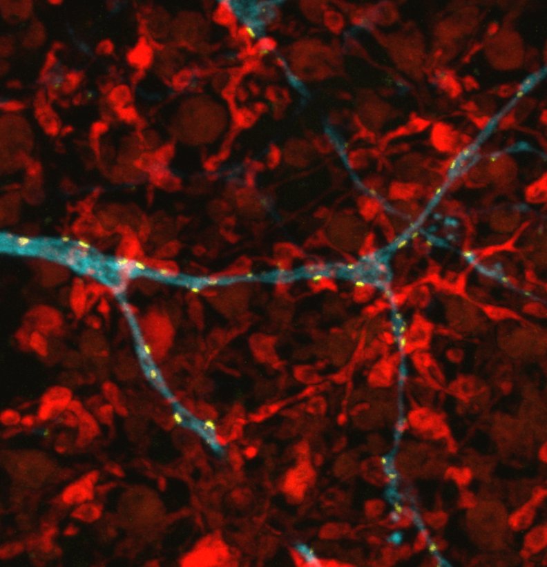
Image by Josh Morgan.
Red: ON bipolar cells terminals. Cyan: Ganglion cell dendrites. Yellow: Postsynaptic density protein PSD95
Rod bipolar cells of the mouse retina.
Image by Luca Della Santina.
Red: Rod bipolar cells. Blue: ON bipolar cells. Green: Goα immunostaining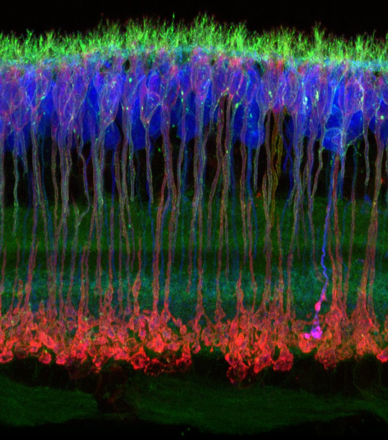
Image by Luca Della Santina.
Red: Rod bipolar cells. Blue: ON bipolar cells. Green: Goα immunostaining
Zebrafish.
Image by Eric Schroeder.
Yellow: Fluorescent protein expression in the Q16 line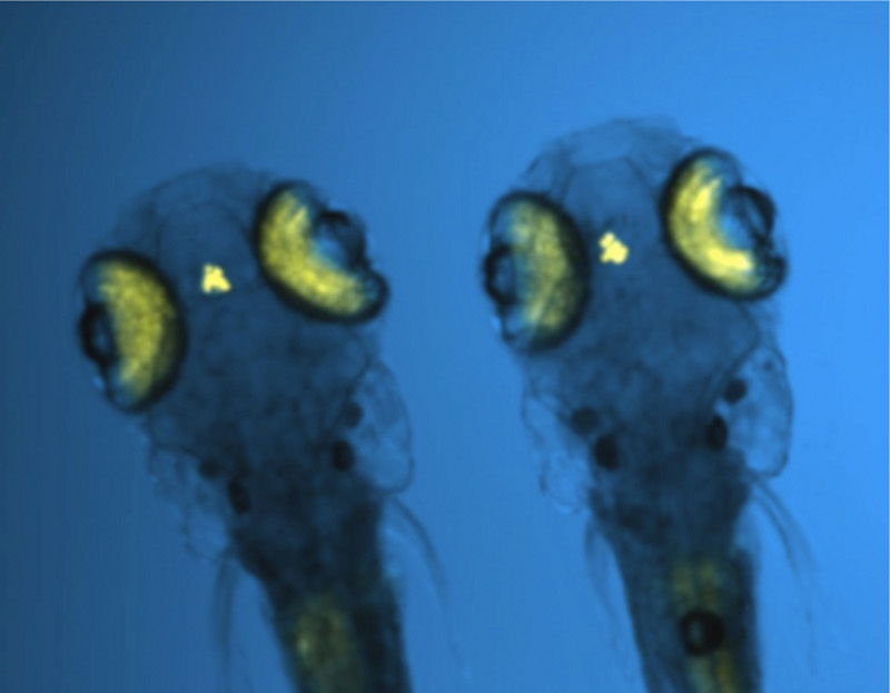
Image by Eric Schroeder.
Yellow: Fluorescent protein expression in the Q16 line
Neuroligin2 distribution on the dendrites of an ON A-type RGC after disruption of GABAergic signaling.
Image by Adam Bleckert.
Blue: Ganglion cell. Green: Neuroligin2. Purple: Remaining GAD67 immunoreactivity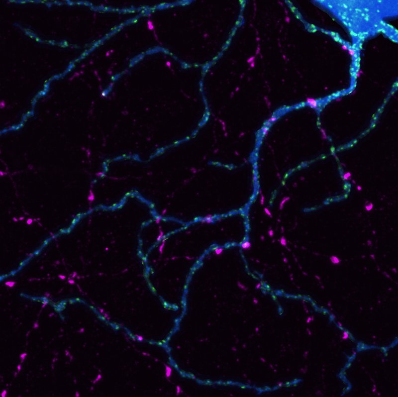
Image by Adam Bleckert.
Blue: Ganglion cell. Green: Neuroligin2. Purple: Remaining GAD67 immunoreactivity
Ribbons within a type 6 mouse cone bipolar cell terminal.
Image by Haruhisa Okawa.
Red: Cone bipolar cell axon terminal. Grey: CtBP2 immunolabeling of ribbons.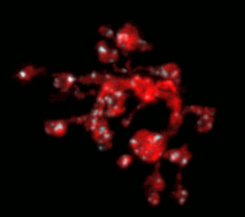
Image by Haruhisa Okawa.
Red: Cone bipolar cell axon terminal. Grey: CtBP2 immunolabeling of ribbons.
Cone mosaic of the zebrafish eye.
Image by Sachihiro Suzuki.
Red: Red cones, Green: Green cones, Cyan: Blue cones. Purple: Ultraviolet cones.
Image by Sachihiro Suzuki.
Red: Red cones, Green: Green cones, Cyan: Blue cones. Purple: Ultraviolet cones.
GABA receptors at the level of mouse rod bipolar cell terminals.
Image by Mrinalini Hoon.
Blue: Rod bipolar cells. Red: GABA receptors. Yellow: GABAergic presynaptic terminals.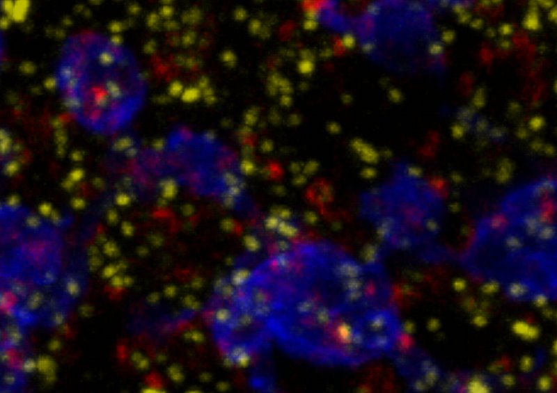
Image by Mrinalini Hoon.
Blue: Rod bipolar cells. Red: GABA receptors. Yellow: GABAergic presynaptic terminals.
Mouse type type 6 cone bipolar cells contacting photoreceptors.
Image by Felice Dunn.
Gray: Cone bipolar cells. Cyan: Cone terminals. Mouse age: 23 days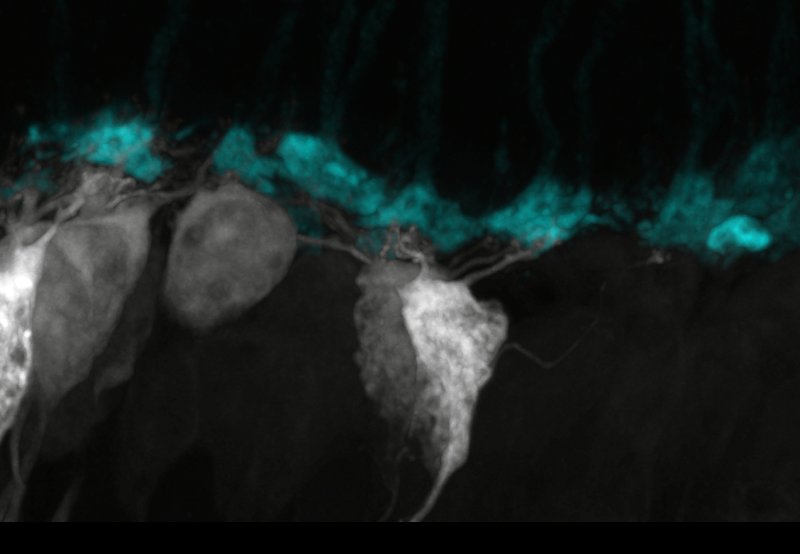
Image by Felice Dunn.
Gray: Cone bipolar cells. Cyan: Cone terminals. Mouse age: 23 days
Image of a live zebrafish
Image by Takeshi Yoshimatsu.
Cyan: Cellular membranes labeled with CFP in the Q01 line at 3 days post fertilization.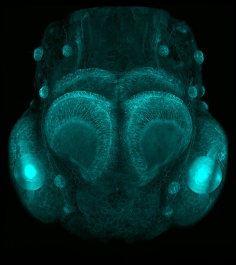
Image by Takeshi Yoshimatsu.
Cyan: Cellular membranes labeled with CFP in the Q01 line at 3 days post fertilization.
Contacts between a mouse cone bipolar cell and ganglion cell.
Image by Haruhisa Okawa.
Red: Bipolar cell. Blue: Ganglion cell. Green: Postsynaptic density protein PSD95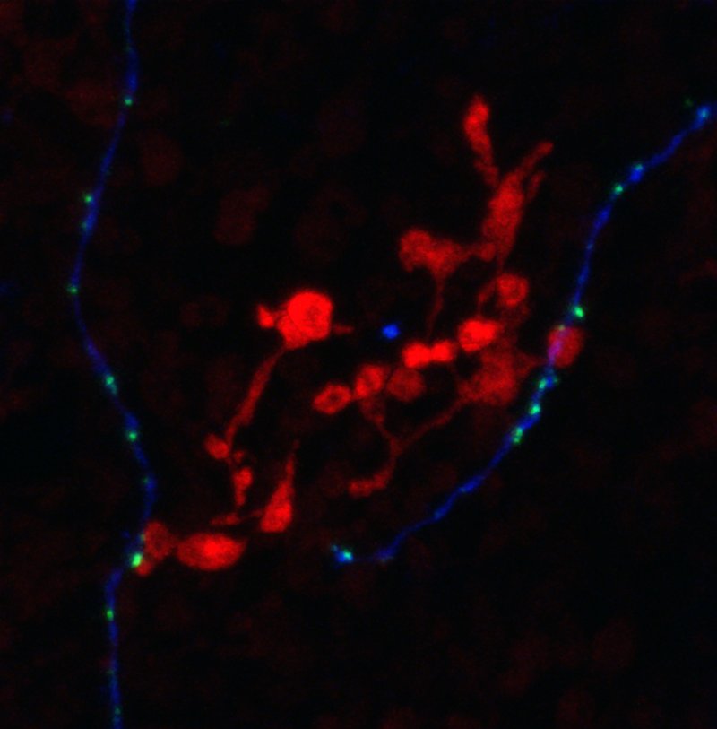
Image by Haruhisa Okawa.
Red: Bipolar cell. Blue: Ganglion cell. Green: Postsynaptic density protein PSD95
Vertical section of the mouse retina.
Image by Josh Morgan.
Purple: Cones. Orange: Horizontal cells. Green: Bipolar cells. Magenta: Amacrine + Ganglion cells.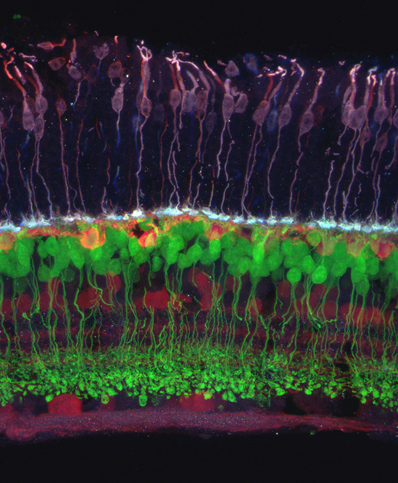
Image by Josh Morgan.
Purple: Cones. Orange: Horizontal cells. Green: Bipolar cells. Magenta: Amacrine + Ganglion cells.
H3 Horizontal cell of zebrafish retina.
Image by Takeshi Yoshimatsu.
Yellow: Horizontal cell. Purple: Ultraviolet cone terminals. Cyan: Blue cone terminals.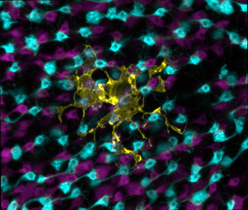
Image by Takeshi Yoshimatsu.
Yellow: Horizontal cell. Purple: Ultraviolet cone terminals. Cyan: Blue cone terminals.
Mosaic of horizontal cells in the early neonatal mouse retina of the GAD1-GFP mouse.
Image by Rachel Huckfeldt.
Individual horizontal cells in the mosaic are pseudocolored.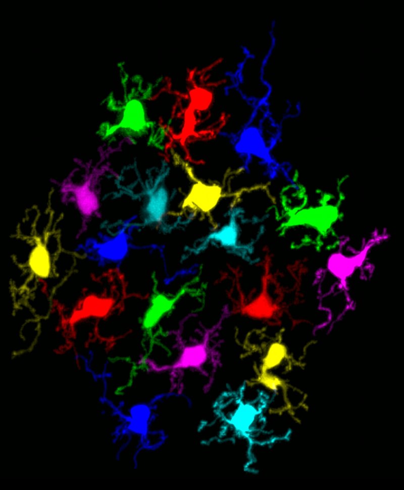
Image by Rachel Huckfeldt.
Individual horizontal cells in the mosaic are pseudocolored.