|
Overall Research
Our lab is interested in neuronal circuit assembly in development, circuit disassembly in degeneration and circuit reassembly upon cellular regeneration. Our studies are based on the vertebrate retina of zebrafish and mice. We apply a diversity of approaches including in vivo and in vitro confocal and multiphoton imaging, electron microscopy, transgenic methods and electrophysiology to investigate neuronal structure, function and connectivity in normal and perturbed retinas. 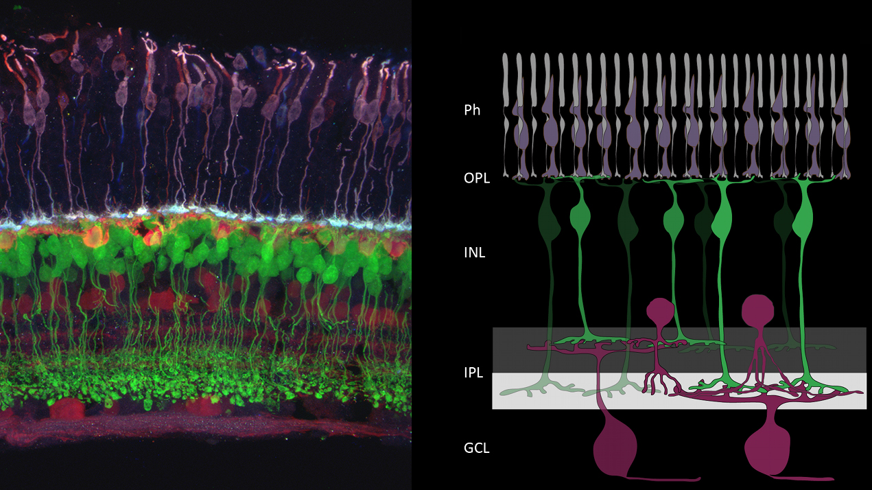
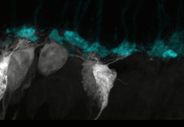
Ph: photoreceptors. OPL/IPL: Outer/Inner Plexiform Layers. INL: Inner Nuclear Layer. GCL: Ganglion Cell Layer. Circuit development in zebrafish 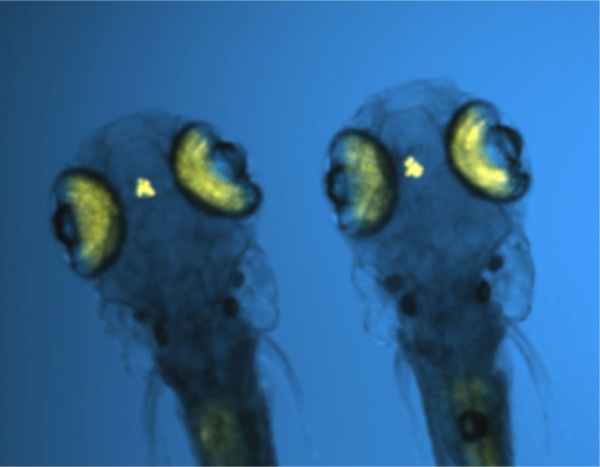
Zebrafish (Danio rerio) develop rapidly and are visually responsive by about 4-5 days postfertilization. Because the embryo and young larvae can be maintained relatively transparent, we are able to track structural changes in zebrafish retinal neurons from the time of their genesis until they form circuits in the living animal. Retinal neurons are visualized by expression of fluorescent protein driven by cell-specific promoters. Labeling is achieved either by injection of DNA at the one-cell stage, or in stable transgenic lines.
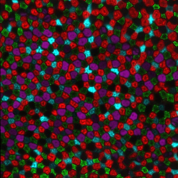
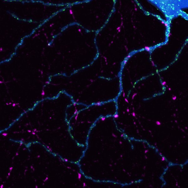
Circuit development in mice By combining biolistic methods, transgenic animals with labeled retinal cell types, and immunohistochemistry, we are reconstructing detailed connectivity maps of neurons in the inner and outer retina. We are interested in the mechanisms that regulate the development of excitatory connections between photoreceptors and bipolar cells and between bipolar cells and retinal ganglion cells. 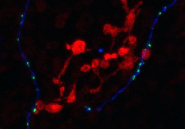
We are also investigating the mechanisms that shape the development of inhibitory connections onto the dendrites and axons of retinal neurons. We are currently focusing on the role of neurotransmission in establishing and maintaining synaptic connectivity between retinal neurons. 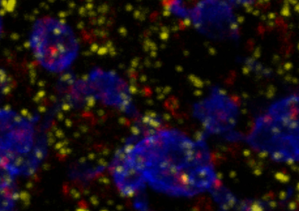
Together with Phil Horner’s lab, we are exploring the steps of neuronal circuit disassembly in a mouse model of glaucoma using multielectrode array recording together with anatomical techniques. The image below shows a piece of retina placed on the 60 electrode array. Using white-noise stimuli, one can generate a movie of the stimulus over time that evoked spikes in a recorded cell (more information on this technique). 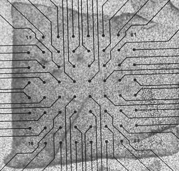
|