
CROSS SECTIONS FROM BLADE TIP TO RHIZOME
"A professor can teach me a term and its definition. A textbook can illustrate this term with a schematic diagram. A field trip can expose me to the ideas behind ecology. A microscope however, allows me to explore a whole new world of science... the rush of not knowing what a cross section might reveal is addictive and the outcome is almost always rewarding."
The following cross sections were made from a specimen of Zostera marina.
All cross section photographs were taken using an Olympus BH-2 Microscope and a QImaging MicroPublisher 3.3 RTV camera.
© Jessica Smith 2008
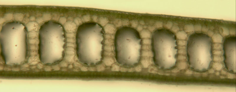
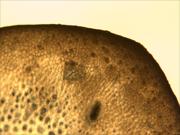
Top section of Z. marina blades |
|
|
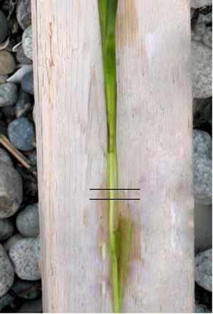
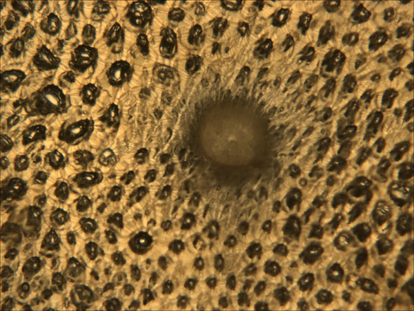
Middle section of Z. marina blade.
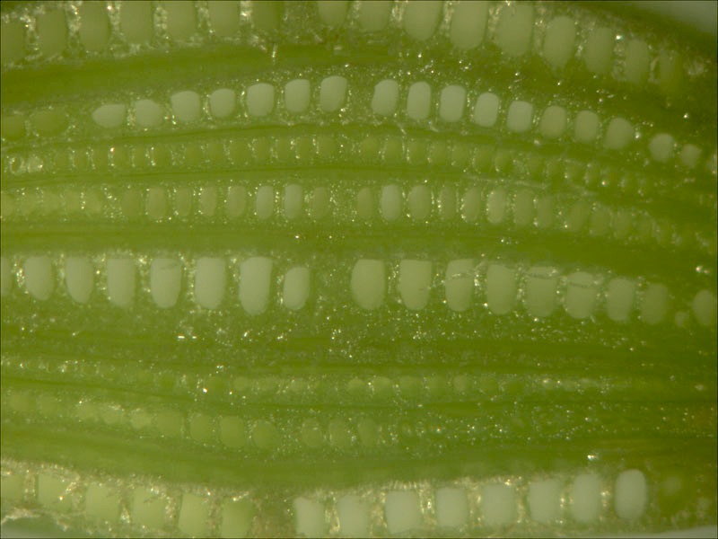
Cross section through the lower region of a Z. marina stem - showing the numerous blade layers, varying in developmental stage.
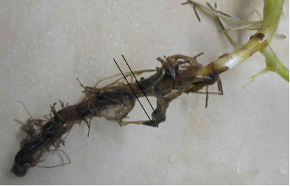
Z. marina rhizome and root system.
Cross section through the rhizome of Z. marina - middle of section (xylem lucunae evident in center).
Cross section through the rhizome of Z. marina - edge of section.