

3D Optical Projection Tomographic Microscopy
Currently diseases are diagnosed using a conventional optical microscope in a pathology or cytology lab after the cells have been fixed and stained. These images from cells on a microscope slide are two-dimensional (2D) from a single perspective view, like an old-fashion chest X-ray. What if these images are three-dimensional like a CT scan used for earlier detection and localization of lung nodules? This advancement is what has been created with the Optical Projection Tomography Microscopy (OPTM) or Cell-CT instrument, (Cell-CT is a trademark of VisionGate Inc. and all movies are Copyright 2008 VisionGate Inc.).
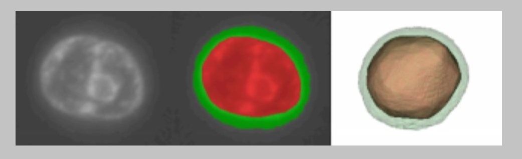
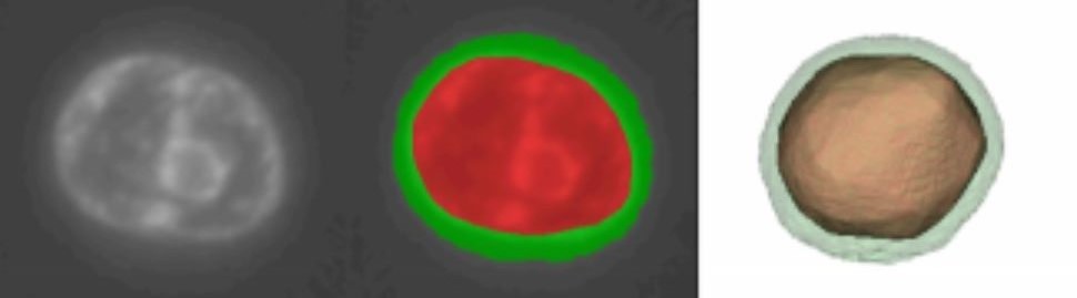
Volumetric (3D) optical imaging of individual cells and nuclei for the earliest detection of cancerous and pre-cancerous conditions, infectious diseases, and effect of drug therapies. In most pathological and cytological analyses, tissue biopsies and cells are imaged in vitro (outside the body) using standard optical microscopes and absorption-based stains. Although cells and nuclei are three-dimensional, this standard imaging technique is only two-dimensional with only one viewing perspective. The development of the Optical Projection Tomography Microscope (OPTM) has allowed 180-degree viewing of individual cells and nuclei at submicron spatial resolution that is isometric. Three-dimensional features are more easily recognized and quantitatively measured using the OPTM, such as the volume, 3D-shape, surface area, surface texture, and 3D features of nuclear invaginations can be used as more sensitive classifiers for earlier conditions of cancer and pre-cancer.
Publications
The following HPL publications are related to this research area:
[bibtex file=HPL_publications.bib group=year group_order=desc key=Agarwal2014a,Agarwal2014,Das2014,Das2014b,Das2014a,Miao2012,Reeves2012,Miao2011,Miao2010a,Miao2010,Miao2010b,Meyer2009,Miao2009a,Miao2009b,Miao2009,Neumann2008,Fauver2005,Fauver2004]