
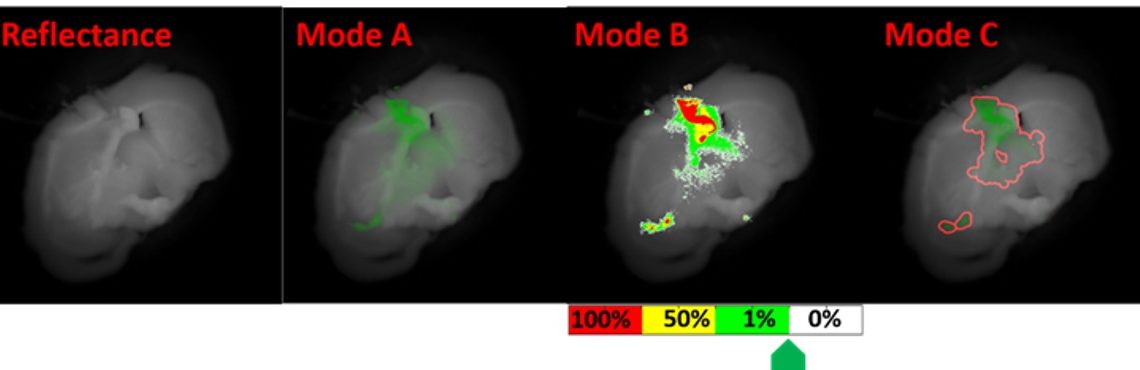
Mode A: enhanced fluorescence overlaid on full color reflectance.
Mode B: delineated tumor regions where colors define percentage of cancer cells in tissue.
Mode C: tunable red boundary detection, where user can select tumor boundary at the % cancer.
References:
Yang Jiang, Emily J. Girard, Fiona Pakiam, Eric J. Seibel, “Ultrathin and flexible 4-channel scope for guiding surgical resections using a near-infrared fluorescence molecular probe for cancer,” Proc. SPIE 10576, Medical Imaging 2018: Image-Guided Procedures, Robotic Interventions, and Modeling, 105762K (13 March 2018); doi: 10.1117/12.2292092
Jiang, Y, Girard, EJ, Pakiam, F, and Seibel, EJ (2019) Calibration of fluorescence imaging for tumor surgical margin delineation: multi-step registration of fluorescence and histological images, Journal of Medical Imaging, 6(2): 025005. doi: 10.1117/1.JMI.6.2.025005
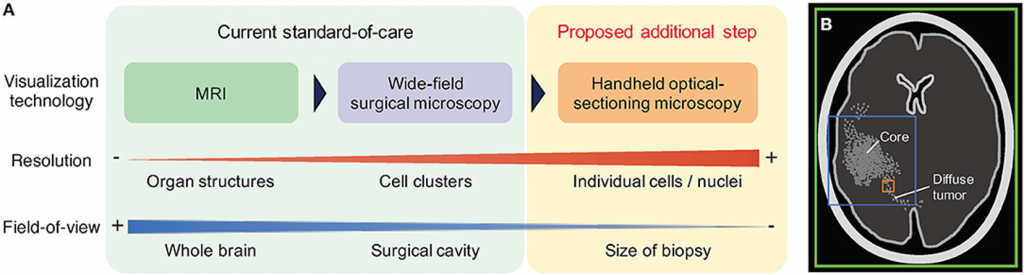
Wei, Roberts, Sanai, Liu (2019) Journal of Neuro-Oncology Fig 1