 |

| Case List | History | Objectives | Radiology | Images | Questions |
| SURGICAL ONCOLOGY Case #1 - A 32 year old with a lesion on her lower... |
|
| Image #6 (#6of 14 found) |
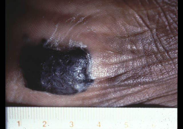
| Nodular melanoma |
| Roll-over to view feature: |
Nodular melanoma (clinical) 
Nodular melanoma (histology)
Melanoma cells
Normal epidermis
Magnification view
Melanoma cells
|
| Detail:
A large collection of malignant melanocytes extending from the epiderms as a solid mass into the mid and deep dermis. This problem represents "vertical growth" of the tumor. Melanoma cells are large with vacuolated cytoplasm. |
 |
| S Taylor © UT Southwestern |
| The Virtual Patient > Case List > SURGICAL ONCOLOGY - Case #1: Images |