 |

| Case List | History | Objectives | Radiology | Images | Questions |
| SURGICAL ONCOLOGY Case #1 - A 32 year old with a lesion on her lower... |
|
| Image #7 (#7of 14 found) |
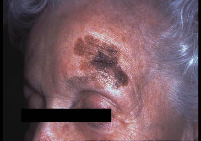
| Lentigo maligna melanoma |

| Detail:
Histology: Groupings or nests of atypical cells in the epidermis with extension into the papillary dermis. Nests coalesce into a sheet of cells that spreads out in a horizontal fashion within the upper levels of the skin. Tumor cells are large with large pleomorphic nuclei and vacuolated cytoplasm. Cells are haphazardly oriented to each other. |
 |
| S Taylor © UT Southwestern |
| The Virtual Patient > Case List > SURGICAL ONCOLOGY - Case #1: Images |