|
Light signaling in the human eye is likely controlled in the outer edge of the retina, UW researchers found in a study published in the Jan. 9, 2003, issue of Nature.
The retina receives light through rods and cones. Cones in the inner retina respond to color. Rods, concentrated on the retina's outer edge, respond to movement with a series of chemical reactions. Rods, like cones, face away from incoming light. Within rods, light causes a chemical reaction with rhodopsin. This begins a chain of stimulation along the visual pathway, which sends information to the brain for interpretation. The brain can detect one photon of light, the smallest unit of energy, when it is absorbed by a photoreceptor.
In the study, atomic-force microscopy of mouse retinae revealed that much of the surface of the membrane was markedly textured with narrow-ruled lines. At high magnification, researchers could see rhodopsin pairs appearing as tidy double rows of protrusions, similar in regularity to eggs in a carton.
This research supports conclusions from previous studies that suggested rod segment disc membranes are densely packed with rhodopsin molecules to allow for optimum absorption of dim light and for subsequent amplification of the faint signals.
The UW researchers from the Department of Ophthalmology were Dr. Yan Liang, senior fellow; Dr. David Saperstein, associate professor; and Dr. Krzysztof Palczewski, Bishop Professor of Ophthalmology who also holds appointments in pharmacology and chemistry. They collaborated with researchers at the M.E. Muller Institute for Microscopy, Biozentrum at the University of Basel, Switzerland and at the International Institute of Molecular and Cell Biology and Department of Chemistry at the University of Warsaw, Poland.
Development Note
The Bishop Foundation has made a significant commitment to vision research at UW Medicine by funding the work of Dr. Krzysztof Palczewski. His studies of light absorption in the eye are revealing the biology of vision. With the assistance of the Bishop Foundation, the Department of Ophthalmology is supporting the basic science research necessary to conceive of new treatments for vision loss.
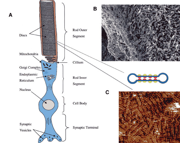
B: Rods in the retina. C: Photons arranged in double rows.
|