|
Using Nanoscience Instrumentation for
Quality Undergraduate Education (unique) in Nanotechnology Undergraduate Education (NUE) |
|
NSF 0634088 |
|||
|
Home
|
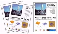
– a one week
hands-on SPM Workshop |
||||
|
Lab
Workbooks
entire books downloadable as PDF (click on year) |
on o
Introduction
to Scanning Force Microscopy o
Scanning
Force Microscopy and Dip-Pen Nanolithography o
AC-Mode
imaging and Electrostatic Force Microscopy |
||||
|
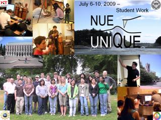
Students will gain
hands-on experience involving a wide variety of nanotechnology/nanoscience
applications, using some of the most versatile nano-tools based on Scanning
Probe Microscopy (SPM). With an intensive one-week schedule and a low student
to instrument and student to TA ratio of 4:1, deep and lasting learning will
occur. The intense 40 hours one-week workshop will provide students with the
opportunity to apply their theoretical knowledge from prior lecture courses. List of Institutions and Disciplines of Past NUE UNIQUE Participants. |
|||||
|
Do you want to take part on the next workshop? |
|
||||
|
Eligibility: Costs: |
To be eligible for the program you
must be: 1. UG student in the second year enrolled
at a 4 year higher educational institution or senior student in a 2 year
higher educational institution (e.g., Community College). Since 2009 we also accept a small number of gifted
graduate students in the first year of their graduate program. 2. Majoring in engineering, materials
science, chemistry, or physics, 3. Available to participate throughout the entire SPM
Workshop. There
is no room for other classes during that week. Successful applicants are
responsible for travel and adequate insurance. This workshop has
been offered for free to all participants thanks to our sponsors, the
National Science Foundation, GEMSEC and Nanosurf GmbH. |
||||
|
Instructors:
R.M. Overney
M. Sarikaya |
Prof. René M Overney (Chem. Prof. Mehmet Sarikaya (Mat. Sci.) is known for his
pioneering efforts and ideas in Molecular
Biomimetics. By merging recent advances in molecular biology and genetics
with state-of-the-art engineering and nanocharacterization from the physical
sciences, his and his collaborators’ goal is to shift the biomimetic
materials science paradigm from imitating Nature to designing materials to
perform artificial nanofunctions. It is the intent to combine Nature’s proven
molecular tools, such as proteins, with synthetic nanoscale constructs to
make molecular biomimetics a full-fledged methodology. To this end, at the
Genetically Engineered Materials Science and |
||||
|
Synopses of Lab Units:
|
Introduction
to Scanning Force Microscopy The
student will become familiar with contact mode Scanning Force Microscopy
(SFM) as an imaging technique and as ultra-sensitive force sensor. Scanning
Force Microscopy and Dip-Pen Nanolithography The
student will become familiar with contact mode Scanning Force Microscopy
(SFM) as an imaging technique, and be introduced with Dip-Pen Nanolithography
(DPN). AC-Mode
imaging and Electrostatic Force Microscopy This lab unit
introduces Electrostatic Force Microscopy to characterize the electrical
properties of a blended conjugated polymer film by studying the changes in
tip oscillation due to electrostatic force gradients between the tip and the
sample. In his
lab unit students are characterizing protein-material using intermittent
non-contact (NC) scanning force microscopy (SFM) in both fluid medium and in
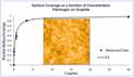
air to quantify surface adsorption. The material analyzed are graphite
adsorbed blood clotting proteins,
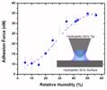
fibrinogen (Fb), to mimic a bio-response to prosthetic heart valve devices. This lab
unit introduces a scanning force microscopy (SFM) based force displacement
(FD) technique, FD analysis, to
study local adhesion, elastic properties, and force interactions between
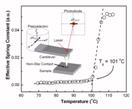
materials. This lab
unit introduces a scanning force microscopy (SFM) based mechanical
(sinusoidal) perturbation method referred to as force modulation microscopy,
to explore thermomechanical properties in polymers around the glass
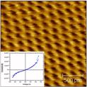
transition. This lab
unit introduces scanning tunneling microscopy (STM) technique, used to obtain
real space atomic resolution images of conductive surfaces. The tunneling
spectroscopy mode of STM is employed to examine local density of state (LDOS)
of the surface. |
||||
|
|
Five laboratory units (including
the Introduction to Scanning Force Microscopy) will be tackled through the
yearly Summer Workshop. The laboratory units are mostly from this list.
Typically one new laboratory unit can be expected every year. |
||||