
| Home |
| 3-D Echo |
| 3-D MRI |
| Applications |
| Quantitative Angiography |
| Centerline™ |
| Visual Guidance |
| Personnel |
| Publications |
| Contact Us |
Contrast Ventriculographic Analysis
Quantitative analysis of left ventricular volume from contrast ventriculograms has been the gold standard against which measurement by other imaging modalities was compared.The most commonly used is the area-length method in which the ventricular contour is compared to an ellipsoid of revolution. The diameter (D) of the ellipse is computed from the area (A) enclosed by the contour and its longest length (L). Volume is calculated using the mean of areas thus computed in each of two orthogonal views. See Fig. 1 for a graphic demonstration of the area-length method, below.
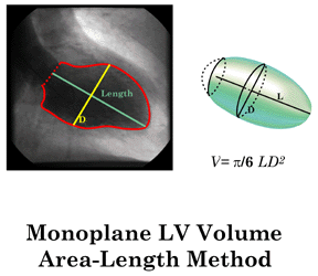 |
Left: RAO view of the LVgram at end diastole, with manually-traced endocardial outline and calculated axial length L and diameter D. Right: LV volume represented as an ellipsoid with major diameter L and minor diameters =D. In this example, only one plane is used. |
A more accurate volume results from use of both RAO and LAO views, and the resultant ellipsoid of minor diameters Drao and Dlao.
The measured volume is corrected for the space occupied by trabeculae and papillary muscles, whose volumes are not measured independently, using a regression equation.
The area-length method has been extensively validated in in vitro hearts as well as in vivo. Measured volumes agree closely with known values (r=0.995, standard error of the estimate =8.2 ml). Accuracy and variability in the application of this method in the clinical setting have been well characterized.
The area-length method is sometimes criticized for comparing the left ventricle to a geometric figure, questioning the relevance of this method to disease-distorted hearts. It should be noted, however, that the method provides accuracy comparable to that of Simpson's rule for measuring the volume of the right ventricle, despite this chamber's irregular shape.
Back to Top