|
|
|
3-D Visual Guidance
Visual anatomic guidance is active computer assistance that visually displays how the image plane moves relative to the target organs anatomy as the examiner manipulates the ultrasound transducer over the patients body. This anatomic information is provided in a real-time graphic display that the examiner can monitor in addition to the image while manipulating the ultrasound transducer. Visual guidance helps the examiner appreciate the spatial location and orientation of the current image plane relative to the anatomic target. Thus visual guidance helps to overcome the most significant disadvantage of ultrasound: the dependence on examiner training and experience.
Studies to date have shown that visual anatomic guidance assists sonographers and relatively inexperienced physician examiners acquire images in the correct anatomic plane, reduces variability in measuring cardiac dimensions, and improves accuracy in measuring right ventricular volume1,2.
Potential applications of visual guidance include:
Improved accuracy and reproducibility in measuring cardiac parameters;
Facilitation of training in ultrasound for sonographers and physicians;
Assistance to rural health care providers with limited training and/or experience;
Facilitation of image-guided procedures such as biopsy;
Guidance for therapeutic ultrasound such as high intensity focused ultrasound against tumors and hemorrhage.
References Cited
1. Dorosz J, Bolson EL, Waiss MS, Sheehan FH.
Three-dimensional visual guidance improves the accuracy of calculating right ventricular volume with two-dimensional echocardiography.
J Am Soc Echocardiogr 2003; (in press).
2. King DL, Harrison MR, King DL Jr, Gopal AS, Martin RP, DeMaria AN. Improved reproducibility of left atrial and left ventricular measurements by guided three-dimensional echocardiography.
J Am Coll Cardiol 2003;20:1238-1245.
 |
|
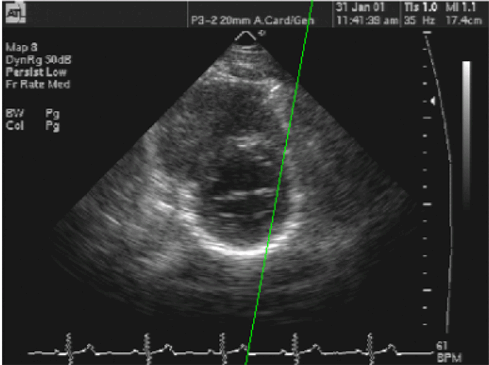
Figure 1. Line of intersection displays in real-time the location of the current image plane (green line) relative to a previously acquired image. However the orientation of the current image plane in the out-of-plane dimension cannot be assessed here. |
 |
|
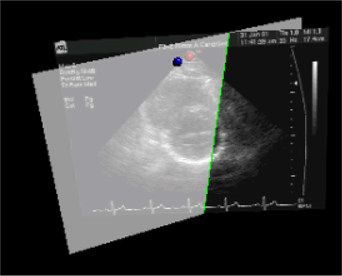
Fig. 2. Visual guidance display of the current image semi-transparently with the scout image. This demonstrates the location and orientation of the current image plane in 3D space relative to the scout image. |
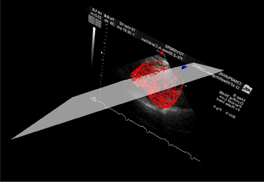 |
| Fig. 3. The most advanced visual guidance display provides a rapidly estimated 3D reconstruction of the left ventricle (red mesh). This illustrates the spatial position and orientation of the current image plane (translucent) much more anatomically. The right ventricle can also be estimated. |
Back
to Top
|