[Skill Modules
>>
Heart Sounds & Murmurs
>>
Techniques
]
Techniques: Heart Sounds & Murmurs
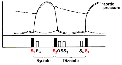
1st & 2nd Heart Sounds
Note: The first heart sound can usually be heard easily with both the bell and the diaphragm but the diaphragm is invaluable for analyzing the second heart sound, with the stethoscope usually best placed at the midleft sternal edge.

back to top
First Heart Sound
Loud Heart Sound
- Hyperdynamic (fever, exercise)
- Mitral stenosis
- Atrial myxoma (rare)
Soft First Sound
- Low cardiac output (rest, heart failure)
- Tachycardia
- Severe mitral reflux (caused by destruction of valve)
Variable Intensity of First Sound
- Atrial fibrillation
- Complete heart block
back to top
Second Heart Sound
Audible expiratory splitting means > 30 msec difference in the timing of
the aortic (A2) and pulmonic (P2) components of the second
heart sound.
- Splitting of S2 is best heard over the 2nd left intercostal space
- The normal P2 is often softer than A2 and rarely audible at apex
- Differential Diagnosis of P2 audible at apex
- Significant pulmonary hypertension
- Atrial septal defect
- Findings should be present in both upright and supine positions: normal
subjects may have expiratory splitting when recumbent that disappears when
upright.
Split S2
| Type |
Inspiration |
Expiration |
Cause |
| Normal or physiologic |
 |
 |
|
| Wide, fixed splitting |
 |
 |
Atrial septal defect |
| Wide split, varies with inspiration |
 |
 |
Pulmonary stenosis, RBBB |
| Paradoxical splitting |
 |
 |
Hypertrophic cardiomyopathy |
Loud Aortic Component of Second Sound
- Systemic hypertension
- Dilated aortic root
Soft Aortic Component of Second Sound
Loud Pulmonary Component of Second Sound
Proceed to the next technique
|


