|
|
[Skill Modules
>>
Heart Sounds & Murmurs
>>
Techniques
]
Techniques: Heart Sounds & Murmurs
Murmurs (general) | Systolic | Diastolic
Systolic Murmurs
| valvular murmurs |
 |
 |
 |
 |
| nonvalvular murmurs |
 |
 |
 |
|
back to top
Causes:
- Blood flow through a structure normally closed during systole (mitral or
tricuspid valves or the interventricular septum).
- Blood flow through a valve normally open in systole but abnormally narrowed
(e.g. aortic or pulmonary stenosis).
- Increased blood flow through a normal valve (a flow murmur).
back to top
Analyze the murmur for
back to top
Where murmurs occur in systole:
back to top
Systolic Murmurs are classified as:
Holosystolic
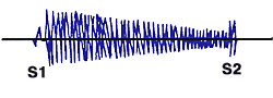
| Holosystolic: Regurgitation across AV valves (mitral and tricuspid) or ventricular septal defect |
| Timing: |
Similar intensity throughout the length of systole |
| Cause: |
Blood flow through an incompetent valve |
| Examples: |
Mitral or tricuspid valve or ventricular septal defect |
Mid or late systolic
| Timing: |
Murmur starts in mid- or late systole |
| Cause: |
Valve is competent at the start of systole
but starts to leak 1/2 way through. |
| Examples: |
Mitral valve prolapse. |
Midsystolic
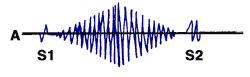
| Midsystolic (crescendo-decrescendo): Aortic stenosis, aortic sclerosis, "flow murmurs," pulmonic stenosis |
| Timing: |
Starts quietly at the beginning of systole, rise to a crescendo in midsystole and then become quiet again towards the end of systole |
| Cause: |
Murmurs that are due to blood being forced through a narrowed |
| Examples: |
Aortic stenosis, aortic sclerosis, "flow murmurs," pulmonic stenosis |
Early Systolic
| Timing |
Aortic stenosis |
MR* |
HCM# |
| Early |
shshssh |
dub |
|
MR when S2heard at base but not apex |
|
| Mid |
lub |
shshsdub |
|
Less likely MR |
|
| Late |
lub |
shshshs
(obliterates S2) |
|
Less likely MR |
|
| Holosystolic |
shshshshshhshshs
(obliterates S1 & S2) |
|
|
|
Where it is best heard and where it radiates to
| Location of Maximal Intensity |
Radiation |
Typical for |
| 2nd right intercostal space |
Right carotid artery |
Aortic stenosis |
| 5th or 6th left intercostal space |
Left anterior axillary line, left axilla |
Mitral regurgitation (including mitral regurgitation due to mitral valve prolapse) |
| Left axilla Lower left sternal border |
LRSB, Epigastrium, 5th ICS mid left thorax |
Tricuspid regurgitation |
| 5th left intercostal space mid- left thorax |
Lower left sternal border |
Hypertrophic cardiomyopathy |
back to top
What it sounds like
| Quality |
Aortic stenosis |
MR* |
HCM# |
| Musical (honk or coo) |
Usually aortic |
|
|
| Nonmusical |
|
|
|
| Blowing |
Usually aortic |
|
|
| Harsh |
|
|
|
back to top
What happens during special maneuvers
Murmur analysis with dynamic auscultation  |
| Maneuvers |
| |
Rt. sided |
Lt. sided |
| TR/PS |
Aortic stenosis (AS) |
MR* |
HCM# |
| Change with respiration |
| |
Inspiration |
 |
Decreases or no change |
|
| To decrease flow |
| |
Valsalva maneuver |
|
 |
|
|
| |
Squat to stand |
|
|
variable |
|
| To increase flow |
| |
Leg elevation |
|
No decrease |
No decrease |
|
| |
Handgrip |
|
|
|
|
| |
Stand to squat |
|
|
variable |
|
*MR=mitral regurgitation
#HCM=hypertrophic cardiomyopathy |
*You can also distinguish between AS and MR by changes in intensity
after changes in cycle length. Listen for a beat after a PVC. With a longer
time between beats, there is increased filling, increased contractility and
decreased afterload. This increases the flow across the mitral valve as more
blood flows forward with the decrease in afterload, decreasing the intensity
of the MR murmur.
|