Category Archives: EEG Sessions

EEG Sessions Journal Club: Experimental manipulation of face-evoked activity in the fusiform gyrus of individuals with ASD

Perlman, SB, Hudac, CM, Pegors, T, Minshew, NJ, and Pelphrey, KA. Experimental manipulation of face-evoked activity in the fusiform gyrus of individuals with Autism. (2011) Soc Neuroscie; 6(1): 22-30.

This month, one of the articles discussed at the EEG Team Journal Club was ‘Experimental manipulation of face-evoked activity in the fusiform gyrus of individuals with Autism’, co-authored by our own Dr. Caitlin Hudac! In this study, researchers used fMRI brain scans and eye gaze manipulation to investigate neural activation in the fusiform gyrus (FFG, a part of the brain specializing in facial recognition) and the amygdala (a part of the limbic system that processes emotional reactions).

Aberrant eye contact is a common feature of Autism Spectrum Disorder (ASD). Eye-tracking experiments have identified atypical gaze patterns among individuals with ASD, specifically in response to social stimuli and faces. Similarly, atypical activation of the fusiform gyrus (FFG), fusiform face area (FFA) and the amygdala in response to social stimuli have been observed in a multitude of fMRI and brain imaging studies comparing ASD and typically developing (TD) participants.

Brain imaging research findings are mixed, however – some studies found a hypoactivation (a smaller response to stimuli) in these regions among the ASD groups, while others found equivalent or even hyperactivation. These discrepant findings may be due in part to the research methods employed in conjunction with underlying neural mechanisms. FFG and amygdala function might be impaired in ASD brains, but perhaps hypoactivation is occurring simply because individuals with autism aren’t looking at eyes or socially salient stimuli. In an attempt to address this posit, Perlman & colleagues manipulated eye gaze to see if FFG and amygdala activation could be normalized in ASD brains when participants were drawn to look at eyes.

12 adults with ASD and 7 TD adults completed an fMRI brain scan while looking at a picture of a fearful male face. (Prior research has demonstrated that fearful faces result in stronger amygdala activation than neutral faces). The fearful face was presented in five different conditions: Nose Fixation, Free Viewing, and Low, Medium, and High Eye Fixation. In the fixation conditions, red crosshairs appeared either on the nose 100% of the time or the eyes for varying lengths of time (Low 32%, Medium 48%, and High 56%). The crosshairs manipulated participants’ eye gaze, such that they drew attention to different areas of the face.

An external file that holds a picture, illustration, etc.
Object name is nihms237852f1.jpg

(Image taken from Perlman’s article)

In line with existing research, TD brains showed more activation in the FFG and amygdala compared to ASD brains during the Free Viewing condition, suggesting that TD participants spent more time looking at the eyes. Interestingly, when eye gaze was manipulated, FFG activation in the ASD brains increased to levels similar to TD brains. In other words, when individuals with ASD are directed to look at eyes, FFG activation is normalized, suggesting that this part of their brain is capable of functioning as a TD brain would function. This was not true of the amygdala, however – eye gaze manipulation had no effect in the ASD brains.

Perlman’s findings have important implications, specifically for treatment and behavioral intervention. Social deficits, including lack of eye contact, can have cascading effects on children with autism. Improved eye contact may have positive effects on early development, learning, and social functioning. As is the case with nearly all research, these findings raise more questions. If the FFG of autistic brains can be normalized through manipulated eye gaze, what is preventing it from activating naturally? When eye gaze was manipulated, why was the FFG normalized but the amygdala remained unaffected?

This article was an interesting read and pretty digestible, even for the ‘non-scientific reader.’ Check it out!

EEG Sessions: EEG Journal Club – Cui, et. al. 2016

EEG Sessions presents ‘EEG Journal Club’, where members of the Bernier Lab’s EEG team read and discuss an article of interest and share their thoughts on our blog.  This month, the EEG team discussed the following article:

Cui, T., Want, P. P., Liu, S., & Zhang, Xin. (2016). P300 amplitude and latency in autism spectrum disorder: a meta-analysis. European Child and Adolescent Psychiatry, 2016, 1-14. doi: 10.1007/s00787-016-0880-z

The Bernier Lab EEG Journal Club recently discussed a research article in which Tingkai Cui and colleagues (2016) performed a meta-analysis on existing event-related potential (ERP) research. A meta-analysis aggregates data from previously published papers to produce robust results for a particular research question or topic. This paper integrated work from 32 studies targeted the P300 component, including data from 407 ASD and 457 neurotypical participants. Participants were presented with frequent auditory stimuli (e.g. a beep), interspersed with infrequent stimuli that differed in tone or pitch. In some experiments, a third, novel sound was included. In some experiments, participants were asked to respond to the infrequent stimuli either motorically (e.g. pressing a button) or mentally (e.g. counting silently). P300 is a neural response occurring around 300 milliseconds after the onset of a novel stimuli. P300 responses can be used to make inferences about attention and decision-making. The P300 component can be further broken down into P3a, which is elicited in the novel tone, no-response conditions, and P3b, which appears when a response to stimuli is requested.

Cui and colleagues conducted a thorough literature review using multiple library databases, selecting studies that met a specific criteria. Inclusion required that studies had both ASD and typically functioning participants and used an ERP technique to report P300 amplitude and/or latency. Cui, et al. calculated and used a standard mean difference to compare the ASD group against the typically functioning group. Cui and colleagues found that participants with ASD had reduced amplitude of the P3b component relative to comparison groups. They found no difference in P3b latency, P3a amplitude, or P3a latency between groups. Reduced amplitude of the P3b among children with ASD suggests that these groups displayed abnormal information processing – but only in tasks where they had to discriminate between and respond to various stimuli. There are a number of ways to interpret the decreased P3b amplitude in the ASD group. For example, perhaps children with ASD have fewer neural resources or perhaps they simply focus their attention on different things. In other words, perhaps  children with ASD focus more on details rather than the ‘bigger picture.’ A reduced P3b amplitude may be a result of how participants in the ASD group allocated their attention.

Journal club reactions…

Brianna: As previously mentioned, one of the hallmark features of autism is its heterogeneity. The children in both ASD and control groups could have varying degrees of abilities. For example, there was no mention of IQ, which could be related to the lack of P3a in ASD groups. Cui pointed out the different paradigms used may yield different P3a components. In future replication studies, it would be wise to select one reliable paradigm that works best and test it on a large sample size. Our journal club was also curious to know if there were any correlations between reaction times and P3b effects. Because some children with autism have motor delay, we wondered if reaction time is the best behavioral measure for this type of experiment.

Caitlin: The P300 component is a very valuable asset for characterizing the attention system in autism for both low- and high-functioning children. I really liked this article because it organized potential reasons why different studies may have variable findings, including whether the experiment activated visual or auditory systems, whether the participant was required make a response, and how often “pop-outs” occur. As a scientist, we spend a lot of time trying to figure out how to build our experiments so that we capture the overall brain process, rather than the brain process only in [a specific scenario]. Although there are not enough similar papers to pick the “best” paradigms (more work to be done!), this paper helped me narrow down possible methods for us to use here in the Bernier lab.

Daisy: The result from this article is concord with past findings that ASD individual usually display an aberrant pattern in P300 component than TD. The main reason I like about article is that it nicely laid out some potential reasons of why results vary from study to study. In consideration of the heterogeneity of ASD symptoms, results vary due to the experiment paradigms they use (e.g. whether it is visual or auditory; whether participants were asked to make a response). P300 component happens rather late in the brain response stage and it usually infers the decision-making process. In the future, I think it will be a valuable information to collect when we work with clinical population in combination of social and behavioral features. A potential question that I am wondering is how P300 serve as an indicator of executive function.

EEG Sessions: An Introduction to EEG

eeg-photo
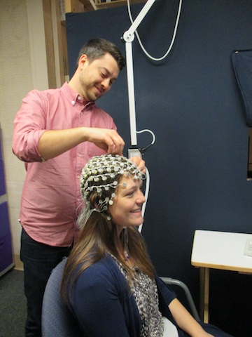
Welcome to the Bernier Lab’s new blog series “EEG Sessions”, where we will share with you EEG related topics, our thoughts on recent papers, and other relevant information from the Bernier Lab.  To start off, we will describe why we measure brain activity using electroencephalography (EEG) and what the data represents.

Here at the Bernier Lab, we research genotypes and phenotypes related to autism spectrum disorder (ASD), which means that we capture a thorough and multifaceted picture of each participant by integrating genetic sequencing,  behavioral assessments, and neurophysiological measures. One of the neurophysiological measures  we use regularly is electroencephalogram (EEG). EEG is a noninvasive procedure that tracks and records brain waves through electrodes that are affixed to the scalp. In our lab, we use an EEG netcap with hundreds of recording sites that is similar in structure to a swimming cap. EEG collects information about brain activity down to the millisecond(!), but it is hard to determine exactly where the brain signals originate within the brain. In other words, EEG provides a very reliable representation of when neural activity occurs, but we have to use discretion to determine where it occurs.

To better understand how brain activity relates to specific aspects of cognitive function, we control what our participants see or hear. Sometimes we show pictures or movies (with or without sounds). Other times we ask participants to sit quietly or close their eyes. We call this method “event-related potentials” (ERP), which refers to the fact that we record brain signals (i.e., potentials) in response to specific stimuli (i.e., events, such as a picture of a face). Stimuli may be visual (e.g. images flashing on a screen) or auditory (e.g. different beeps, tones, and sounds). We examine ERP responses in milliseconds and refer to specific parts of the brain activity, or brain waves, as ‘components.’ Components of interest are measured both in latency (When does the brain response occur?) and amplitude (How strong is the response?). There are predictable brain responses that occur at specific time points, often with expected amplitudes. In our ERP analyses, we measure selected components and compare them across different groups (e.g. children with ASD versus typically developing children).

In conducting EEG experiments, we hope to establish specific biomarkers that may aid in the diagnoses of autism. Currently, ASD can only be diagnosed through a series of clinical assessments. While these measures are reliable, they are built for toddlers and older children, simply due to the nature of the activities involved. Trying to diagnose children who are nonverbal or have cognitive impairments is especially challenging. Biomarkers could aid with diagnosis of children who are difficult to evaluate with traditional clinical ASD measures, including infants. This is especially significant because early intervention is critical and effective. If, for example, we found that a group of children with ASD share a brain signature that is different from other groups of children with ASD, we could use that knowledge to aid in early diagnosis and intervention. Designated biomarkers that aid early diagnosis of autism could also provide insight about anticipated behaviors and challenges for individual children. Such knowledge would be valuable in determining which treatments and therapies would be most beneficial for that child.

Establishing biomarkers for autism is no easy task. We know that ASD is a complex, multifaceted disorder, and as such, there will always be a number of factors that are difficult or impossible to address. Isolating ERPs can also prove challenging due to inherent brain differences associated with chronological age and developmental stage. Genetic events, comorbid disorders, and the heterogeneity of ASD further add to the complexity of establishing reliable biomarkers of autism. As a result, it is not uncommon for research to have mixed or contradictory findings. This is certainly true in EEG/ERP research. One of the goals of our EEG journal club is to think critically about existing research in order to improve our own methods and produce reliable findings with clinical applications.