
Summary of the article Factors associated with the improvement of vocal fold movement: an analysis of LEMG and laryngeal CT parameters. Mengsteab PY, Kwon JY, Han TR, Kwon TK, Kim DH, Kim SJ. J Electromyogr Kinesiol. 2015 Feb;25(1):1-7. doi: 10.1016/j.jelekin.2014.08.004. Epub 2014 Aug 27. PMID: 25217204 Clinical Trial. https://pubmed.ncbi.nlm.nih.gov/25217204/
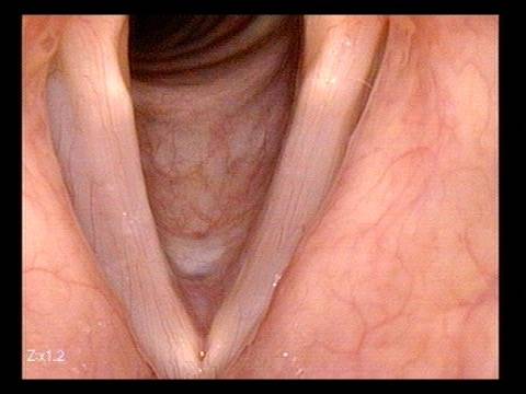
Laryngeal nerve injury occurs when there is damage to one of the nerves that helps the larynx function. The damage can be caused by injury, tumor, infection, or surgery. Patients may present symptoms of a hoarse voice, difficulty swallowing, or loss of voice.
The injury/damage can be diagnosed using a tool called laryngeal electromyography (LEMG) that measures the electrical activity of the muscles in the larynx, also known as the voice box. However, the interpretation of the results obtained with this tool can be distorted by muscle contracture, weakness, or atrophy. Therefore, the prediction of a favorable prognosis using this tool is inaccurate.
Another diagnostic method commonly used to study the larynx is an imaging tool called computed tomography (CT). Scans or images of the cross-section of the larynx are obtained using X-rays. Imaging can often reveal subtle muscle and nerve changes because it can scan deeper structures.
This study compared these two diagnostic tools to determine if both would improve the prognosis of patients with recurrent laryngeal nerve injury.
The researchers found that CT allows for better detection of muscle atrophy compared to LEMG.
Through the analysis of images obtained with CT, it was possible to observe an improvement of the larynx muscles through regeneration and determine a more accurate prognosis than the one obtained with LEMG alone.
These results are important because they show that pairing both techniques will help to give a better, more accurate prognosis, to patients who suffer from recurrent larynx injury.