
MRI stands for Magnetic Resonance Imaging. An MRI scanner uses a magnetic field and radio pulses to create 3-dimensional pictures of tissue. In our studies, we look at two basic types of MRI images: structural and functional.
 |
Structural:
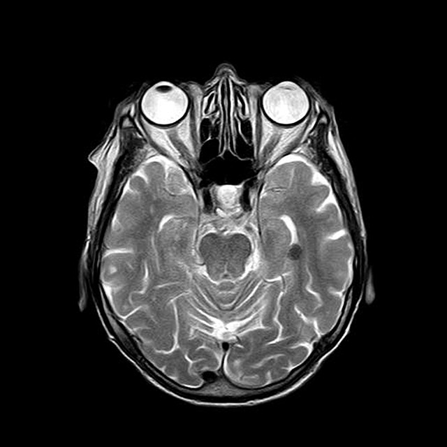
T1 and T2 Weighted: T1 and T2 weighted are both structural scans that produce a 3D image of the brain using either spin echo or gradient echo sequences. The black/white color of T2 is the reverse of that of T1. DTI: Diffusion Tensor Imaging (DTI) is a type of structural scan that is able to detect the diffusion of water molecules in tissue. |
Theoretically, this shows us the white matter fiber tracts of the brain. The basic idea behind this is that water molecules moving along the axon will move more quickly than those moving across or in other directions.
Functional:
Functional MRI (fMRI) looks at oxygen consumption in various regions of the brain during certain time blocks. These time blocks correlate to specific tasks performed in the scanner. Regions with higher levels of oxygen build-up are believed to be more active.
What to expect:


When participants come to Seattle Children’s to get an MRI for one of our studies, they get to wear special goggles so that they don’t see the inside of the MRI machine. They also wear earplugs and headphones to help block out the noise. For parts of the scan, participants get to watch their favorite movie and for other parts, they will complete tasks in which they respond using a button box. We just ask participants to lay down, relax, and stay still!