[Skill Modules
>>
Thyroid
>>
Techniques
]
Techniques: Thryroid Exam
 There are several physical examination maneuvers described for examination of the thyroid described below that are at least moderately sensitive and specific. Much of the exam is based on physiological reasoning and tradition rather than on studies of reliability or precision. Combining the examination and association signs and symptoms increases the accuracy of the physical examination of the thyroid. There are several physical examination maneuvers described for examination of the thyroid described below that are at least moderately sensitive and specific. Much of the exam is based on physiological reasoning and tradition rather than on studies of reliability or precision. Combining the examination and association signs and symptoms increases the accuracy of the physical examination of the thyroid.
Goiter: Examination of the thyroid for size
Note: An enlarged thyroid is referred to as a goiter. There is no direct correlation between size and function- a person with a goiter can be euthyroid, hypo- or hyperthyroid.
A normal thyroid is estimated to be 10 grams with an upper limit of 20 grams or 2 to 4 teaspoons.
Examination for goiter can increase the possibility of thyroid disease in patients with symptoms of hypo- or hyperthyroidism, in determining the choice of treatment in hyperthyroidism and monitoring the response to therapy directed at decreasing the size of the thyroid in cases of symptomatic goiter.
The examination consists of three portions:
- Inspection,
- Palpation, and
- Synthesis of data from these techniques
In addition to palpating for size, also note the gland texture, mobility, tenderness and the presence of nodules.
Inspection
Inspection: Anterior Approach
- The patient should be seated or standing in a comfortable position with the neck in a neutral or slightly extended position.
- Cross-lighting increases shadows, improving the detection of masses.
- To enhance visualization of the thyroid, you can:
- Extending the neck, which stretches overlying tissues
- Have the patient swallow a sip of water, watching for the upward movement of the thyroid gland.
 251KB video demo from Return to the Bedside 251KB video demo from Return to the Bedside
Inspection: Lateral Approach
- After completing anterior inspection of the thyroid, observe the neck from the side.
- Estimate the smooth, straight contour from the cricoid cartilage to the suprasternal notch.
- Measure any prominence beyond this imagined contour, using a ruler placed in the area of prominence.
Palpation
Note: There is no data comparing palpation using the anterior approach to the posterior approach so examiners should use the approach that they find most comfortable.
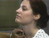
Palpation: Anterior Approach
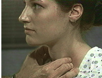
- The patient is examined in the seated or standing position.
- Attempt to locate the thyroid isthmus by palpating between the cricoid cartilage and the suprasternal notch.
- Use one hand to slightly retract the sternocleidomastoid muscle while using the other to palpate the thyroid.
- Have the patient swallow a sip of water as you palpate, feeling for the upward movement of the thyroid gland.
- 454KB video demo from Return to the Bedside.
Palpation: Posterior Approach
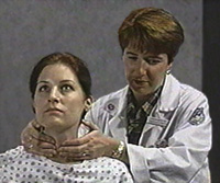
- The patient is examined in the seated or standing position.
- Standing behind the patient, attempt to locate the thyroid isthmus by palpating between the cricoid cartilage and the suprasternal notch.
- Move your hands laterally to try to feel under the sternocleidomstoids for the fullness of the thyroid.
- Have the patient swallow a sip of water as you palpate, feeling for the upward movement of the thyroid gland.
Note: This traditional technique is based on physiological reasoning; data of effectiveness is lacking.
Synthesis of data from these techniques
Using the data from anterior and lateral inspection and from palpation, categorize the gland as:
- goiter ruled out [normal or small (1 to 2 times normal) with lateral prominence <2 mm ],
- goiter ruled in [large (& 2 times normal) or lateral prominence >2 mm] or
- inconclusive.
See Evidence Base and Differential Diagnosis.
back to top
Nodules: Examination of the thyroid for nodularity
Thyroid nodules are common (prevalence 4%). Half of the thyroids glands examined by ultrasound or direct visualization (surgery or autopsy) have nodules. Physical examination detects approximately 10% of the nodules found by these methods. Nodules increase in frequency with age and are four times more likely in women than men. Less than 5% of all nodules are cancerous.
Technique
- The location of the thyroid is identified by inspection.
- Using the anterior or posterior approach, palpate the thyroid to identify nodules
- Note the size and number of nodules.
- Note the consistency of the nodule.
- Palpate regional lymph nodes for consistency and mobility.
- Take a look at a teaching demo video.
|