Contributed by: Steven J. Rockoff, MD and Diana L. Lam, MD – June 1, 2020
Question 1
A 40-year-old woman presents for a baseline screening mammogram:
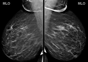
You note a large mass in the left breast. What is the best description for the location of this mass?
A. 1:00 (upper outer quadrant)
B. 1:00 (upper inner quadrant)
C. 5:00 (lower outer quadrant)
D. 5:00 (lower inner quadrant)
E. 11:00 (upper inner quadrant)
F. 11:00 (upper outer quadrant)
Answer
A. 1:00 (upper outer quadrant)
Explanation: To interpret a mammogram, one must become familiar with the proper way to describe the location of a finding such as a mass or calcifications. Standard reporting should include the following:
- Laterality (left or right breast)
- Quadrant (upper outer, upper inner, lower outer, or lower inner)
- O’clock face (i.e. 12:00, 1:00, 2:00, etc…)
O’clock face is determined by visualizing that you are standing in front of the patient, looking at the breast with a clock superimposed upon it. Note that any o’clock position will not be in the same quadrant of each breast (i.e. a 2:00 mass in the right breast is upper inner quadrant, but a 2:00 mass in the left breast is upper outer quadrant).
As per the ACR BI-RADS manual, other location descriptors that can be used are:
- Depth (anterior third, middle third, or posterior third)
- Distance from the nipple
- A few terms that can be used in lieu of a quadrant: “central”, “retroareolar”, “axillary tail”
Determining the quadrant and o’clock face of a finding requires being familiar with the standard display of a mammogram. If you are not yet familiar with the orientation of a mammogram, see the below annotations:


Question 2
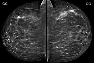
What is your assessment and recommendation after reading the screening mammogram?
A. BI-RADS 0 (Incomplete); Recommend diagnostic ultrasound
B. BI-RADS 1 (Negative); Recommend one year follow-up
C. BI-RADS 2 (Benign); Recommend one year follow-up
D. BI-RADS 3 (Probably Benign); Recommend six month follow-up
E. BI-RADS 4 (Suspicious); Recommend biopsy
Answer
A. BI-RADS 0 (Incomplete); Recommend diagnostic ultrasound
Explanation: This is the patient’s first (baseline) mammogram and there is a left breast mass in the upper outer quadrant that needs further characterization before deciding final management. Targeted diagnostic ultrasound is the correct next step.
Question 3
The ultrasound of the expected position of the mass is performed. A representative image:
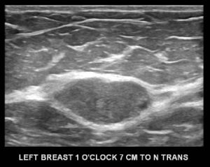
What is your assessment and recommendation?
A. BI-RADS 0 (Incomplete); Recommend diagnostic MRI
B. BI-RADS 1 (Negative); One year follow-up
C. BI-RADS 2 (Benign); One year follow-up
D. BI-RADS 3 (Probably Benign); Six month follow-up
E. BI-RADS 4 (Suspicious); Ultrasound-guided biopsy
F. BI-RADS 5 (Highly Suspicious); Ultrasound-guided biopsy
Answer
D. BI-RADS 3 (Probably Benign); Six month follow-up
Explanation: This non-palpable, solid, circumscribed oval mass can be assessed as “Probably Benign”, meaning that it has a less than a 2% chance of being malignant. The assessment of BI-RADS 3 can be given with a recommendation for short term follow-up (diagnostic ultrasound in six months). Once two years of stability has been documented for this mass, it can finally be designated benign with no specific follow-up needed.
As per the ACR BI-RADS manual 5th edition, there are three mammographic findings which can be designated as BI-RADS 3, Probably Benign:
- A solid mass which is non-palpable, non-calcified, circumscribed, oval or round (as seen in this case).
- A non-palpable focal asymmetry with no ultrasound correlate.
- A solitary group of punctate calcifications.
Question 4
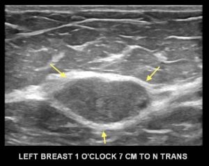
Maybe you were wondering about the smaller mammographic mass that is posterior to the dominant mass. Here is the ultrasound of that second mass:

What is this mass?
A. Cyst
B. Hamartoma
C. Lymph Node
Answer
C. Lymph Node
Explanation: This is the normal sonographic appearance of an intramammary lymph node, with a thin hypoechoic cortex and a hyperechoic fatty hilum.