Skip to content

Case 19

Contributed by: Steven J. Rockoff, MD and Diana L. Lam, MD – June 1, 2020

Question 1

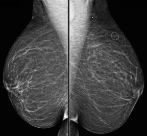
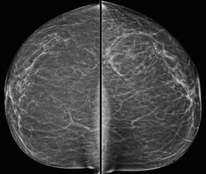
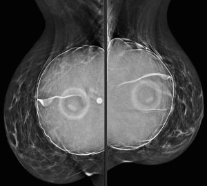
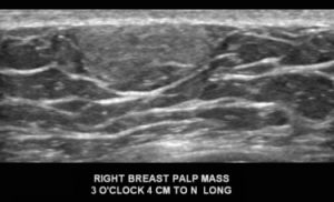
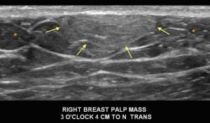
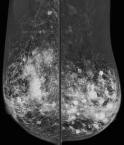
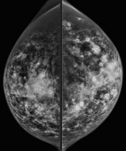
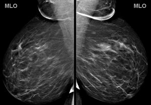
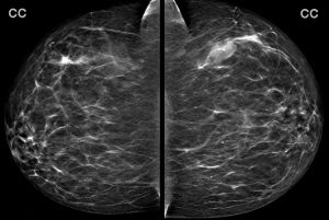
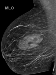
A 43-year-old woman presents for a screening mammogram:

What is the most striking finding?

A. Right axillary lymphadenopathy
B. Marked vascular calcifications
C. Diffuse skin thickening
D. Left nipple inversion
E. Multiple bilateral round and oval massses

Answer

E. Multiple bilateral round and oval massses

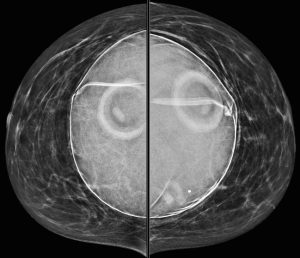
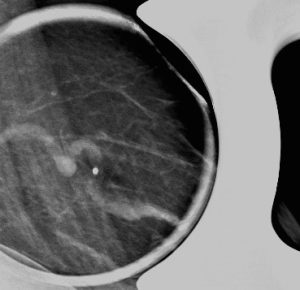
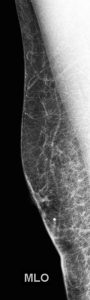
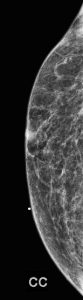
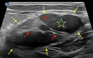
Explanation: There are multiple circumscribed round and oval masses (arrows on the below images). At least two masses are seen in each breast on these 2D images (when scrolling through the tomosynthesis images, even more masses can be seen).

There are no enlarged axillary lymph nodes. There are several skin folds projecting over the axilla on the right MLO image.

There is no nipple inversion or retraction. The left nipple looks like it is behind the skin on the CC image, however this is due to the nipple not being in profile when the image was acquired. Both nipples appear normal and symmetric on the MLO projections.

Question 2

What is your assessment and recommendation?

A. BI-RADS 0 (Incomplete); Recommend diagnostic mammogram and ultrasound
B. BI-RADS 1 (Negative); Recommend one year follow-up
C. BI-RADS 2 (Benign); Recommend one year follow-up
D. BI-RADS 3 (Probably Benign); Recommend six month follow-up
E. BI-RADS 4 (Suspicious); Recommend biopsy

Answer

C. BI-RADS 2 (Benign); Recommend one year follow-up

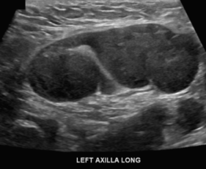
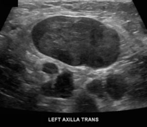
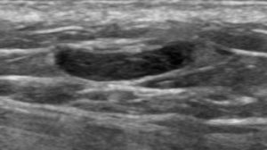
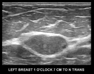
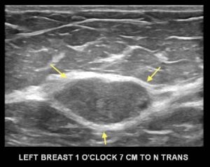
Explanation: There are multiple, bilateral, round and oval, circumscribed, benign-appearing masses. This is a situation for which there is strong data to suggest that there is an essentially 0% likelihood of malignancy. The multiplicity and bilaterality is indicative of multiple cysts, or less often fibroadenomas.

If there are at least three similar benign-appearing masses, with at least one in each breast, then they can be designated benign (BI-RADS 2). No further diagnostic work-up is indicated unless one of the masses appears significantly different from the others or is growing significantly.

In women who have these multiple benign-appearing masses, it can be normal for them to slightly fluctuate in size between yearly exams, as cysts are known to do.

Case 18

Contributed by: Steven J. Rockoff, MD and Diana L. Lam, MD – June 1, 2020

Question 1

A 71-year-old woman presents with a palpable lump. A BB skin marker is placed over the area of concern and a diagnostic mammogram is performed.


What is the dominant abnormality?
A. Enlarged axillary lymph node
B. Sebaceous cyst
C. Lipoma
D. Supernumerary nipple

Answer

A. Enlarged axillary lymph node

Explanation: The metallic BB marker overlies the axilla and is only seen on the MLO projection. On that image, we see a definitely enlarged lymph node and a second lymph node inferiorly that is also probably enlarged.

Question 2

The mammogram is reviewed, and besides the abnormal lymph nodes, no other suspicious finding is seen in the breasts. An ultrasound is performed of the axilla:


What is your assessment and recommendation?
A. BI-RADS 0 (Incomplete); Recommend diagnostic MRI
B. BI-RADS 1 (Negative); Recommend lymph node biopsy
C. BI-RADS 3 (Probably Benign); Recommend six month follow-up
D. BI-RADS 4 (Suspicious); Recommend lymph node biopsy

Answer

D. BI-RADS 4 (Suspicious); Recommend lymph node biopsy

Explanation: There are multiple abnormal lymph nodes in the left axilla, and so far no evidence of any abnormality in the left breast. The presence of unilateral axillary lymphadenopathy must be presumed to be metastatic breast cancer until proven otherwise. These findings must be deemed suspicious and biopsy of one of the lymph nodes should be performed. Whenever possible, the BI-RADS assessment and recommendation should be concordant.

Question 3

Ultrasound-guided biopsy of a left axillary lymph node yielded metastatic carcinoma, consistent with a primary breast origin.

What is the most appropriate next step?
A. Refer to surgeon for axillary excision
B. Left breast skin punch biopsy
C. Diagnostic MRI
D. PET/CT

Answer

C. Diagnostic MRI

Explanation: In a case of metastatic breast cancer diagnosed by lymph node biopsy, in which the site of the primary tumor cannot be determined by mammography, the most appropriate test to perform next is MRI, which has an extremely high sensitivity for detecting in-situ and invasive breast cancer.

PET/CT is usually only performed in select cases of newly diagnosed known breast cancers when there is a higher likelihood of more advanced locoregional or metastatic spread.

In this patient’s case, MRI was able to identify the site of the primary left breast cancer.

Case 17

Contributed by: Steven J. Rockoff, MD and Diana L. Lam, MD – June 1, 2020

Question 1

A 46-year-old woman presents with a tender and palpable left breast abnormality. A diagnostic mammogram is performed:

What kind of breast implants does the patient have?
A. Prepectoral saline implants
B. Retropectoral saline implants
C. Prepectoral silicone implants
D. Retropectoral silicone implants

Answer

B. Retropectoral saline implants

Explanation: These implants are filled with saline. Note the “see through” density of the saline, compared to the extremely dense silicone implant that was demonstrated in a previous case. We can also see the implant valves and mild rippling of the external implant contour, which are features typically associated with saline implants.

On the MLO images, we can see the pectoralis major wrapping around the anterior portion of the implant, which indicates that they are retropectoral implants (also known as “subpectoral”).

Question 2

The spot magnification mammogram view and targeted ultrasound images that were performed at the site of palpable abnormality are shown here.


What is your diagnosis?
A. Abscess
B. Superficial venous thrombophlebitis
C. Phlegmon
D. Filariasis

Answer

B. Superficial venous thrombophlebitis (Mondor’s disease of the breast)

Explanation: Thrombophlebitis presenting as a partial or total thrombosis of a superficial vein in the breast is colloquially known as Mondor’s disease.  Patients present with a cord-like palpable mass, often with local tenderness and skin erythema. Although the process is most commonly idiopathic, it can also be due to trauma or iatrogenic causes, or rarely, local breast cancer. Unless there is an identifiable ongoing cause, the condition is usually benign and self-limiting, and should be treated supportively with warm compresses and anti-inflammatory medication.

On our mammogram and ultrasound images, we can see the dilated and tortuous affected vessel. Although the vessel is abnormally hypoechoic under ultrasound due to the presence of thrombus, the presence of some color Doppler flow within the vessel indicates partial patency of the lumen.

There is no evidence of abscess or phlegmon. Filariasis is the most common parasitic infection of the breast, which presents with a lump or swelling, and serpiginous calcifications seen on mammography (this is rare in the United States).

Case 16

Contributed by: Steven J. Rockoff, MD and Diana L. Lam, MD – June 1, 2020

Question 1

A 58-year-old man presents with a palpable lump in his right breast.
What is the most appropriate imaging test to perform first?
A. Diagnostic ultrasound
B. Diagnostic mammogram
C. Screening mammogram

Answer

B. Diagnostic mammogram

Explanation: According to the American College of Radiology Appropriateness Criteria, for a male who is 25 years or older, the most appropriate imaging exam to perform first with a clinically indeterminate palpable breast mass is a diagnostic mammogram.

This patient has focal symptoms, so a screening exam is not appropriate. There are also currently no indications to screen males for breast cancer.

A bilateral diagnostic mammogram was performed. Images of the right breast shown here do not demonstrate any mammographic abnormality under the BB skin marker:

Question 2


With the mammogram appearing negative, a diagnostic ultrasound was performed. What is the diagnosis?
A. Cyst; BI-RADS 2 (Benign)
B. Invasive ductal carcinoma; BI-RADS 4 (Suspicious)
C. Abscess; BI-RADS 2 (Benign)
D. Lipoma; BI-RADS 2 (Benign)

Answer

Explanation: This is a circumscribed, oval, hyperechoic mass, which is consistent with a lipoma. They have the same imaging appearance in males and females. They may appear as circumscribed fat-containing masses on the mammogram, particularly if the patient has a lot of fibroglandular tissue. Since this male’s breast is composed predominantly of fat density, the lipoma cannot be easily discerned on the mammogram. On ultrasound, a lipoma (arrows) is either isoechoic or slightly hyperechoic to the surrounding subcutaneous fat (*). Lipomas are benign and when correctly identified on imaging, they do not require further follow-up.

D. Lipoma; BI-RADS 2 (Benign)

Case 15

Contributed by: Steven J. Rockoff, MD and Diana L. Lam, MD – June 1, 2020

Question 1

A 15-year old girl visits her doctor because she is concerned about a right breast mass that seems to be quickly growing.

What is the most appropriate test for her doctor to order?
A. Diagnostic ultrasound
B. Diagnostic mammogram
C. Diagnostic tomosynthesis
D. Diagnostic MRI

Answer

A. Diagnostic ultrasound

Explanation: For any female under the age of 30 presenting with a presenting breast mass, the first imaging exam that should be obtained is a diagnostic ultrasound.  Due to the increased risk of ionizing radiation, the increased likelihood of extremely dense tissue, and the low incidence of breast cancer in this demographic, mammography is not the appropriate first imaging exam to utilize in this situation.

Question 2

The ultrasound was performed. Representative images of the dominant abnormality are shown here:

This mass measures up to 65 mm in longest axis. What is the most likely diagnosis?
A. Granular cell tumor
B. Juvenile fibroadenoma
C. Hemangioma
D. Invasive ductal carcinoma

Answer

B. Juvenile fibroadenoma

Explanation: Fibroadenoma, a benign fibroepithelial lesion, is the most common breast mass in the pediatric population. Juvenile fibroadenoma, also known as giant fibroadenoma, is a variant characterized by rapid growth to a size of 5 cm or larger. Like normal fibroadenomas, they are usually circumscribed round or oval masses. Biopsy is usually indicated because the potentially malignant Phyllodes tumor can have an identical imaging appearance. Because of their rapid growth and their potential to cause local deformity, they are usually treated with surgical excision.

The other answer choices are possible pediatric breast masses, but are not as common as fibroadenoma.

Case 14

Contributed by: Steven J. Rockoff, MD and Diana L. Lam, MD – June 1, 2020

Question 1

A 40-year-old woman is recalled from her screening mammogram to work-up calcifications in the right breast. Diagnostic magnification views (CC & ML) are obtained:

What kind of calcifications are these? (try zooming in on your browser if needed)

A. Dystrophic
B. Popcorn-like
C. Rim
D. Skin
E. Milk of calcium

Answer

E. Milk of calcium

Explanation: These calcifications appear amorphous on the CC projection, and have a semilunar or crescent-shaped morphology on the 90º lateral (LM) projection. This is the classic appearance of “milk of calcium”, which are sedimented calcifications within cysts. These are benign.

On the CC projection, they milk of calcium often does not have a discrete shape. When x-rays pass through in the perpendicular (lateral) direction, we can see the calcifications layering within the dependent portion of their cysts, creating the characteristic curvilinear or “tea cup” appearance.

Case 13

Contributed by: Steven J. Rockoff, MD and Diana L. Lam, MD – June 1, 2020

Question 1

A 60-year-old woman presents for a screening mammogram:

What is responsible for the appearance of this mammogram?
A. Extremely dense breast tissue with multiple oil cysts
B. History of silicone implant rupture
C. History of direct silicone injections
D. Steatocystoma multiplex

Answer

C. History of direct silicone injections

Explanation: Direct injection of silicone is a method of breast augmentation that is illegal in the United States, but may still be practiced in other countries. The practice results in a mammogram with multiple high density masses (with rim calcifications) and areas of greatly increased density. Extremely dense lymph nodes may also be present. These high-density silicone breast masses make it difficult to detect a breast cancer on both physical exam and mammography.

Steatocystoma multiplex is a skin disorder, manifesting in the breast as multiple bilateral subcutaneous oil cysts. Although this patient has multiple rim calcifications (which can also occur with oil cysts), steatocystoma multiplex would not explain the presence of so many areas of greatly increased density.

Case 12

Contributed by: Steven J. Rockoff, MD and Diana L. Lam, MD – June 1, 2020

Question 1

A 40-year-old woman presents for a baseline screening mammogram:

You note a large mass in the left breast. What is the best description for the location of this mass?
A. 1:00 (upper outer quadrant)
B. 1:00 (upper inner quadrant)
C. 5:00 (lower outer quadrant)
D. 5:00 (lower inner quadrant)
E. 11:00 (upper inner quadrant)
F. 11:00 (upper outer quadrant)

Answer

A. 1:00 (upper outer quadrant)

Explanation: To interpret a mammogram, one must become familiar with the proper way to describe the location of a finding such as a mass or calcifications. Standard reporting should include the following:

  • Laterality (left or right breast)
  • Quadrant (upper outer, upper inner, lower outer, or lower inner)
  • O’clock face (i.e. 12:00, 1:00, 2:00, etc…)

O’clock face is determined by visualizing that you are standing in front of the patient, looking at the breast with a clock superimposed upon it. Note that any o’clock position will not be in the same quadrant of each breast (i.e. a 2:00 mass in the right breast is upper inner quadrant, but a 2:00 mass in the left breast is upper outer quadrant).

As per the ACR BI-RADS manual, other location descriptors that can be used are:

  • Depth (anterior third, middle third, or posterior third)
  • Distance from the nipple
  • A few terms that can be used in lieu of a quadrant: “central”, “retroareolar”, “axillary tail”

Determining the quadrant and o’clock face of a finding requires being familiar with the standard display of a mammogram. If you are not yet familiar with the orientation of a mammogram, see the below annotations:

Question 2

What is your assessment and recommendation after reading the screening mammogram?
A. BI-RADS 0 (Incomplete); Recommend diagnostic ultrasound
B. BI-RADS 1 (Negative); Recommend one year follow-up
C. BI-RADS 2 (Benign); Recommend one year follow-up
D. BI-RADS 3 (Probably Benign); Recommend six month follow-up
E. BI-RADS 4 (Suspicious); Recommend biopsy

Answer

A. BI-RADS 0 (Incomplete); Recommend diagnostic ultrasound

Explanation: This is the patient’s first (baseline) mammogram and there is a left breast mass in the upper outer quadrant that needs further characterization before deciding final management. Targeted diagnostic ultrasound is the correct next step.

Question 3

The ultrasound of the expected position of the mass is performed. A representative image:

What is your assessment and recommendation?
A. BI-RADS 0 (Incomplete); Recommend diagnostic MRI
B. BI-RADS 1 (Negative); One year follow-up
C. BI-RADS 2 (Benign); One year follow-up
D. BI-RADS 3 (Probably Benign); Six month follow-up
E. BI-RADS 4 (Suspicious); Ultrasound-guided biopsy
F. BI-RADS 5 (Highly Suspicious); Ultrasound-guided biopsy

Answer

D. BI-RADS 3 (Probably Benign); Six month follow-up

Explanation: This non-palpable, solid, circumscribed oval mass can be assessed as “Probably Benign”, meaning that it has a less than a 2% chance of being malignant. The assessment of BI-RADS 3 can be given with a recommendation for short term follow-up (diagnostic ultrasound in six months). Once two years of stability has been documented for this mass, it can finally be designated benign with no specific follow-up needed.

As per the ACR BI-RADS manual 5th edition, there are three mammographic findings which can be designated as BI-RADS 3, Probably Benign:

  1. A solid mass which is non-palpable, non-calcified, circumscribed, oval or round (as seen in this case).
  2. A non-palpable focal asymmetry with no ultrasound correlate.
  3. A solitary group of punctate calcifications.

Question 4

Maybe you were wondering about the smaller mammographic mass that is posterior to the dominant mass. Here is the ultrasound of that second mass:

What is this mass?
A. Cyst
B. Hamartoma
C. Lymph Node

Answer

C. Lymph Node

Explanation: This is the normal sonographic appearance of an intramammary lymph node, with a thin hypoechoic cortex and a hyperechoic fatty hilum.

Case 11

Contributed by: Steven J. Rockoff, MD and Diana L. Lam, MD – June 1, 2020

Question 1

A 61-year-old man presents to his doctor because he is concerned about a palpable finding in his right breast. His doctor agrees that he is able to appreciate the area of concern on physical exam.

The most appropriate test to order is:
A. CT of the chest
B. Diagnostic ultrasound
C. Diagnostic mammogram
D. Diagnostic MRI
E. CA-125 blood test

Answer

C. Diagnostic mammogram

Explanation: According to the American College of Radiology Appropriateness Criteria, for a male who is 25 years or older, the most appropriate imaging exam to perform first with a clinically indeterminate palpable breast mass is a diagnostic mammogram.

Question 2

The diagnostic mammogram is performed. A BB skin marker is placed over the area of palpable concern in the right breast.

What is your assessment and recommendation?
A. BI-RADS 0 (Incomplete); Recommend diagnostic ultrasound
B. BI-RADS 1 (Negative); Recommend one year follow-up
C. BI-RADS 2 (Benign); No follow-up needed
D. BI-RADS 3 (Probably Benign); Recommend six month follow-up
E. BI-RADS 4 (Suspicious); Recommend biopsy

Answer

C. BI-RADS 2 (Benign); No follow-up needed

Explanation: This patient has gynecomastia, which is the most common etiology of a tender and palpable breast mass in males. The mammographic appearance of gynecomastia can vary based on the time course, but the classic appearance is increased tissue density in the subareolar region, often with a “flame shape”. Gynecomastia is a benign process of epithelial hyperplasia, and when identified on mammography, does not warrant further work-up. Gynecomastia is not significantly associated with an elevated risk of male breast cancer. In older males, mammography is the first line modality in the setting of a male breast complaint, in part because gynecomastia usually has a suspicious sonographic appearance, and the diagnosis can often be made with mammography alone.

Any suggestion of a mass on mammography, however, should prompt further evaluation with ultrasound and possible ultrasound-guided biopsy.

This patient actually has mild gynecomastia on the left side as well. Bilateral (and asymmetric) gynecomastia is not uncommon.

Case 10

Contributed by: Steven J. Rockoff, MD and Diana L. Lam, MD – June 1, 2020

Question 1

A 37-year-old woman presents for evaluation of a right breast mass that was incidentally seen on a CT exam for an unrelated issue.

Diagnostic ultrasound:

Diagnostic mammogram:

What is your assessment?
A. BI-RADS 0 (Incomplete); Recommend diagnostic MRI
B. BI-RADS 1 (Negative); Recommend one year follow-up
C. BI-RADS 2 (Benign); Recommend one year follow-up
D. BI-RADS 3 (Probably Benign); Recommend six month follow-up
E. BI-RADS 4 (Suspicious); Recommend biopsy

Answer

Explanation: The large mass in the right breast at 9:00 is a breast hamartoma, also known as a fibroadenolipoma. This benign mass has the characteristic “breast within a breast” appearance on mammogram, which is a well-circumscribed oval mass composed of normal fibroglandular and adipose tissue. These are benign malformations and do not require biopsy.

The yellows arrows in these ultrasound images demarcate the capsule of the hamartoma. The green stars denote the hypoechoic areas of fibroglandular tissue within the hamartoma and the red arrows denote the hyperechoic areas of fat within the hamartoma:

Although not needed to make the diagnosis, the CT on which the breast hamartoma was partially visualized also demonstrates multiple internal fat elements within the mass: