Contributed by: Steven J. Rockoff, MD and Diana L. Lam, MD – June 1, 2020
Question 1
A 71-year-old woman presents with a palpable lump. A BB skin marker is placed over the area of concern and a diagnostic mammogram is performed.
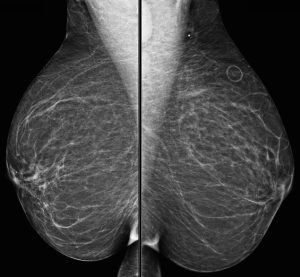
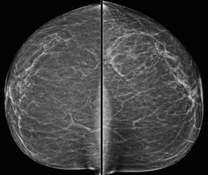
What is the dominant abnormality?
A. Enlarged axillary lymph node
B. Sebaceous cyst
C. Lipoma
D. Supernumerary nipple
Answer
A. Enlarged axillary lymph node
Explanation: The metallic BB marker overlies the axilla and is only seen on the MLO projection. On that image, we see a definitely enlarged lymph node and a second lymph node inferiorly that is also probably enlarged.
Question 2
The mammogram is reviewed, and besides the abnormal lymph nodes, no other suspicious finding is seen in the breasts. An ultrasound is performed of the axilla:
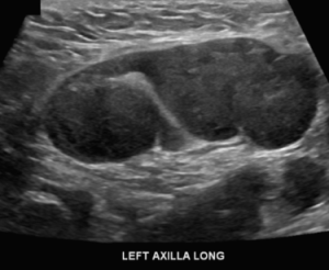
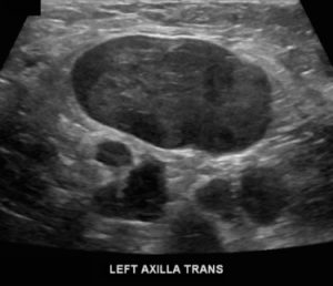
What is your assessment and recommendation?
A. BI-RADS 0 (Incomplete); Recommend diagnostic MRI
B. BI-RADS 1 (Negative); Recommend lymph node biopsy
C. BI-RADS 3 (Probably Benign); Recommend six month follow-up
D. BI-RADS 4 (Suspicious); Recommend lymph node biopsy
Answer
D. BI-RADS 4 (Suspicious); Recommend lymph node biopsy
Explanation: There are multiple abnormal lymph nodes in the left axilla, and so far no evidence of any abnormality in the left breast. The presence of unilateral axillary lymphadenopathy must be presumed to be metastatic breast cancer until proven otherwise. These findings must be deemed suspicious and biopsy of one of the lymph nodes should be performed. Whenever possible, the BI-RADS assessment and recommendation should be concordant.
Question 3
Ultrasound-guided biopsy of a left axillary lymph node yielded metastatic carcinoma, consistent with a primary breast origin.
What is the most appropriate next step?
A. Refer to surgeon for axillary excision
B. Left breast skin punch biopsy
C. Diagnostic MRI
D. PET/CT
Answer
C. Diagnostic MRI
Explanation: In a case of metastatic breast cancer diagnosed by lymph node biopsy, in which the site of the primary tumor cannot be determined by mammography, the most appropriate test to perform next is MRI, which has an extremely high sensitivity for detecting in-situ and invasive breast cancer.
PET/CT is usually only performed in select cases of newly diagnosed known breast cancers when there is a higher likelihood of more advanced locoregional or metastatic spread.
In this patient’s case, MRI was able to identify the site of the primary left breast cancer.