Skip to content

Case 6

Contributed by: Steven J. Rockoff, MD and Diana L. Lam, MD – June 1, 2020

Question 1

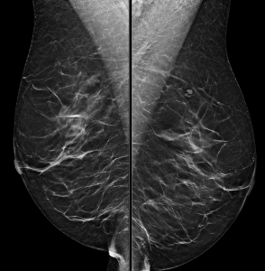
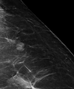
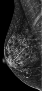
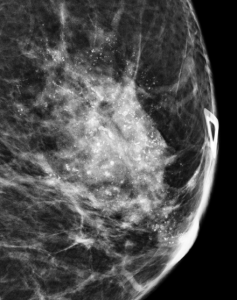
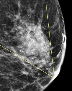
Magnification views were performed for the right breast calcifications seen on a screening mammogram:

What is the best descriptor for this type of calcification?

A. Large rod-like calcification
B. Amorphous calcification
C. Skin calcification
D. Milk of calcium
E. Popcorn-like calcification

Answer

E. Popcorn-like calcification

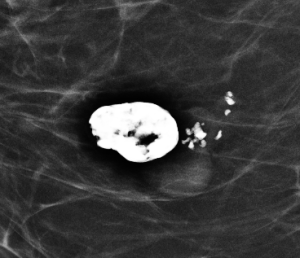
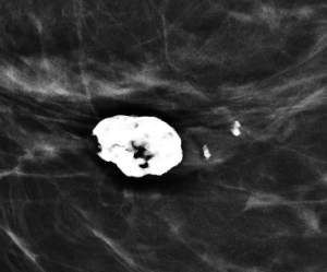
Explanation: These are the large, coalescent, coarse or “popcorn-like” calcifications that are typical of an involuting fibroadenoma.

There are actually probably two or three adjacent fibroadenomas in this location. The dominant large calcification represents the end stage of an entirely calcified involuting fibroadenoma. The smaller adjacent popcorn-like calcifications represent an earlier stage of involution in a different fibroadenoma.

Question 2

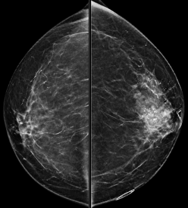
What is the best BI-RADS assessment for this finding?

A. BI-RADS 0 (Incomplete); Recommend diagnostic ultrasound
B. BI-RADS 2 (Benign); Recommend one year follow-up
C. BI-RADS 3 (Probably Benign); Recommend six month follow-up
D. BI-RADS 4 (Suspicious); Recommend biopsy

Answer

B. BI-RADS 2 (Benign); Recommend one year follow-up

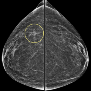
Explanation: A mass with this type of popcorn-like calcifications is almost certainly a fibroadenoma and benign. Having prior mammograms which demonstrate little or no change over time can increase your confidence.

In this patient’s case, there is gradual involution (and slowly increasing calcifications) of this fibroadenoma (image on the left) compared to mammogram five years prior (image on the right):

Case 5

Contributed by: Steven J. Rockoff, MD and Diana L. Lam, MD – June 1, 2020

Question 1

A 48-year-old woman presents for screening mammogram:

How do you best describe the patient’s breast density?

A. The breasts are almost entirely fatty
B. There are scattered areas of fibroglandular density
C. The breasts are heterogeneously dense
D. The breasts are extremely dense

Answer

Either B. There are scattered areas of fibroglandular density, or C. The breasts are heterogeneously dense

Explanation: One of the first steps in interpreting a mammogram is assessing the breast composition, also known as breast density. There are four acceptable categories described in the BI-RADS manual, which are listed above. Breast density is a subjective assessment of how much fibroglandular tissue compared to fat tissue is present in the breast, and also has a correlation to how difficult it may be to interpret a patient’s mammogram. Woman who have “dense” breasts (heterogeneously or extremely dense) have a slightly increased risk of developing breast cancer compared to women with non-dense breasts. This patient’s breasts were reported as being composed of scattered areas of fibroglandular density. Since this is a subjective determination, describing them as heterogeneously dense could also be acceptable.

Question 2

A mass is present in the upper outer quadrant of the left breast:

How is this mass best described?

A. BI-RADS 0 (Incomplete); Recommend diagnostic mammogram and ultrasound
B. BI-RADS 2 (Benign); Recommend one year follow-up
C. BI-RADS 3 (Probably Benign); Recommend six month follow-up
D. BI-RADS 4 (Suspicious); Recommend biopsy

Answer

B. BI-RADS 2 (Benign); Recommend one year follow-up

Explanation: This is the classic appearance of an intramammary lymph node on mammography. This is a normal breast structure, typically appearing as a circumscribed small mass on the mammogram with a lucent notch that corresponds to the anatomic fatty hilum. The most common location is in the upper outer quadrant. If the appearance is not quite classic, a history of stability on prior mammograms is reassuring that this is most likely a normal/benign lymph node.


In this patient’s case, her intramammary lymph node had been unchanged for many years and has the expected appearance of a lymph node on this post-contrast MRI image:

Case 4

Contributed by: Steven J. Rockoff, MD and Diana L. Lam, MD – June 1, 2020

Question 1

A 56-year-old woman presents for a screening mammogram:

How do you best describe the patient’s breast density?

A. The breasts are almost entirely fatty

B. There are scattered areas of fibroglandular density

C. The breasts are heterogeneously dense

D. The breasts are extremely dense

Answer

C. The breasts are heterogeneously dense

Explanation: One of the first steps in interpreting a mammogram is assessing the breast composition, also known as breast density. There are four acceptable categories described in the ACR BI-RADS manual, which are listed above. This patient’s breasts are heterogeneously dense on mammogram, which may obscure small masses.

Question 2

A representative zoomed in image is shown here:

What is the dominant type of calcification that is present?

A. Fine pleomorphic calcifications

B. Large rod-like calcifications

C. Vascular calcifications

D. Linear calcifications

Answer

C. Vascular calcifications

Explanation: These are benign vascular calcifications, present in a diffuse distribution bilaterally. These have the characteristic “tram-track” appearance.

Note that “linear” is not a type of calcification like the other answer choices, but rather is a descriptor for the distribution of calcifications.

Question 3

Although difficult to appreciate on the 2-D whole breast images provided above, there was a screen-detected mass in the lower inner quadrant of the right breast. Here is the mass noted on MLO and spot tomosynthesis CC images:

What is the best descriptor for the margins of this mass on mammogram?

A. Circumscribed

B. Obscured

C. Microlobulated

D. Indistinct

E. Spiculated

Answer

E. Spiculated

Explanation: The numerous thin lines radiating outward from the mass are indicative of spiculated margins, which is a suspicious finding.

A mass with circumscribed margins has at least 75% of its margins sharply demarcated and separable from the surrounding tissue.

A mass with obscured margins has at least 25% of its margin hidden by superimposed or adjacent breast tissue.

A mass with microlobulated margins has multiple small outward undulations.

A mass with indistinct margins has no clear demarcation of its margin from the surrounding tissue.

Question 4

Targeted ultrasound was performed, with a representative image of the mass shown here:

What is the best descriptor for the margins of this mass on ultrasound?

A. Indistinct

B. Angular

C. Microlobulated

D. Spiculated

Answer

A. Indistinct

Explanation: This mass is not circumscribed on ultrasound. The margin between the majority of the mass and surrounding tissue is not clearly defined, and there is a lack of other features that would be seen with the other margin descriptors. This mass’s sonographic margin is best described as indistinct.


Ultrasound-guided biopsy was performed, which yielded invasive ductal carcinoma.

Case 3

Contributed by: Steven J. Rockoff, MD and Diana L. Lam, MD – June 1, 2020

Question 1

A bilateral diagnostic mammogram is performed for a 56-year-old woman with a left breast palpable abnormality and pain:

Other than these whole breast views, what additional views would you like to see in order to make your assessment of the dominant abnormality?

A. Tangential views
B. Spot Magnification views
C. Cleavage views
D. Eklund views

Answer

B. Spot Magnification views

Explanation: There are highly suspicious calcifications in the left breast. In order to best assess the calcifications, spot magnification views are needed, which will allow for a better description of the calcifications’ morphology and distribution.

Tangential views are used to determine whether a mammographic finding is located in the skin, such as skin calcifications.

Cleavage views are used to view the more medial breast tissue of both breasts.

Eklund views, also known as implant-displaced views, are obtained in order to view more breast tissue in women who have breast implants.

Question 2

A spot magnification view in the ML projection is obtained:

How do you describe the distribution and morphology of the calcifications?

A. Grouped distribution; Coarse heterogeneous morphology
B. Grouped distribution; Fine pleomorphic morphology
C. Segmental distribution; Coarse heterogeneous morphology
D. Diffuse distribution; Amorphous morphology

Answer

C. Segmental distribution; Coarse heterogeneous morphology

Explanation: These calcifications are in a segmental distribution, which is a roughly triangular shape which points towards the nipple. This pattern suggests that the calcifications are primarily located within the breast’s ductal system. The individual calcifications predominantly have discrete irregular shapes, are slightly coalescent, and are slightly larger than the fine pleomorphic type of calcification.

Question 3

What is your assessment and recommendation?

A. BI-RADS 0 (Incomplete); Diagnostic US
B. BI-RADS 2 (Benign); One year follow-up
C. BI-RADS 3 (Probably Benign); Six month follow-up
D. BI-RADS 4 (Suspicious); Stereotactic biopsy

Answer

Either D. BI-RADS 4 (Suspicious) or E. BI-RADS 5 (Highly Suggestive of Malignancy); Stereotactic biopsy

Explanation: Either BI-RADS 4 or BI-RADS 5 could be an acceptable assessment for this patient. Stereotactic biopsy is usually the best way to obtain reliable tissue sampling in the setting of suspicious calcifications. You may have noticed the large focal asymmetry which is associated with the calcifications; in such a case, diagnostic ultrasound to look for a target that could be biopsied with ultrasound guidance would also be a reasonable option.

Question 4

You decided to give the left breast BI-RADS 5 in your report. Stereotactic biopsy was performed. There are a large amount of calcifications in the biopsy specimen. The pathology report states that there are fibrocystic changes and other benign findings.

What would you say for radiology-pathology concordance in the addendum to the biopsy report? 

A. Discordant biopsy; Recommend surgical excision
B. Discordant biopsy; Recommend six month follow-up
C. Concordant biopsy; Recommend surgical excision
D. Concordant biopsy; Recommend six month follow-up

Answer

A. Discordant biopsy; Recommend surgical excision

Explanation: 

Segmental distribution has the highest likelihood of malignancy out of all of the calcification distribution descriptors, and coarse heterogeneous morphology is also considered suspicious. Coupled with the fact that there is a focal asymmetry associated with the calcifications, BI-RADS 5 is appropriate. If a BI-RADS 5 (Highly Suggestive of Malignancy) assessment is made, you must be prepared to recommend re-biopsy or surgical excision if the pathology report of the initial biopsy only describes benign findings.

A concordant biopsy means that the pathology findings are a reasonable explanation for the imaging findings. A discordant biopsy (as in this situation) means that the benign pathology findings do not adequately explain the suspicious imaging findings.

In reality, biopsy of this patient yielded invasive ductal carcinoma with ductal carcinoma in-situ (DCIS).

Case 2

Contributed by: Steven J. Rockoff, MD and Diana L. Lam, MD – June 1, 2020

Question 1

A 71-year-old woman presents for a screening mammogram:

What is the best interpretation of this exam?

A. Mass in the right breast
B. Mass in the left breast
C. Focal asymmetry in the right breast
D. Focal asymmetry in the left breast

Answer

C. Focal asymmetry in the right breast

Explanation: According to the ACR BI-RADS Atlas, a focal asymmetry is a unilateral deposit of fibroglandular tissue that does not meet the definition of a mass. It is seen on two projections (CC and MLO), has concave-outward borders, and is usually interspersed with fat. A focal asymmetry that is new or larger since the previous exam (as was the case for this patient) could also be called a “developing asymmetry”.

Question 2

Now that we’ve identified the right breast developing asymmetry, what is the next step?

A. Diagnostic mammogram
B. Diagnostic ultrasound
C. Stereotactic biopsy
D. Follow-up mammogram in six months

Answer

A. Diagnostic mammogram

Explanation: The screening exam would be designated BI-RADS 0, Incomplete. A unilateral right diagnostic mammogram is the correct next step in order to determine if this finding is suspicious enough to warrant biopsy.

Question 3

The right diagnostic mammogram was performed:

The developing asymmetry persists. A targeted ultrasound at the expected position of the mammographic abnormality in the upper outer quadrant is negative. What your assessment and recommendation?

A. BI-RADS 4 (Suspicious); Stereotactic biopsy
B. BI-RADS 3 (Probably Benign); Follow-up mammogram in six months
C. BI-RADS 4 (Suspicious); Diagnostic MRI
D. BI-RADS 4 (Suspicious); Biopsy the approximate area under ultrasound

Answer

A. BI-RADS 4 (Suspicious); Stereotactic biopsy

Explanation: A new and persistent suspicious asymmetry, focal asymmetry, or developing asymmetry that has no definite sonographic correlate should be biopsied with stereotactic guidance. In this case, stereotactic biopsy revealed invasive lobular carcinoma.

Case 1

Contributed by: Steven J. Rockoff, MD and Diana L. Lam, MD – June 1, 2020

Question 1

An 82-year-old woman presents for a screening mammogram:

What is the dominant abnormality?

A. Mass in the right breast
B. Mass in the left breast
C. Focal asymmetry in the right breast
D. Focal asymmetry in the left breast
E. No abnormality

Answer

B. Mass in the left breast

Explanation: According to the ACR BI-RADS Atlas, a mass is a three dimensional, space-occupying finding that is seen on two different mammographic projections. It has convex-outward borders and is usually dense in the center compared to its periphery.

Question 2

What is your assessment and recommendation?

A. BI-RADS 4 (Suspicious); Ultrasound-guided biopsy
B. BI-RADS 4 (Suspicious); Stereotactic biopsy
C. BI-RADS 0 (Incomplete); Diagnostic MRI
D. BI-RADS 0 (Incomplete); Diagnostic mammogram and ultrasound

Answer

D. BI-RADS 0 (Incomplete); Diagnostic mammogram and ultrasound

Explanation: One must become familiar with the BI-RADS Assessment Categories and their corresponding management recommendations. The next step in the work-up of a screen-detected, potentially suspicious mass is diagnostic mammography and ultrasound. In this situation, BI-RADS Assessment Category 0 (“Need Additional Imaging Evaluation”) should be assigned and the patient is recalled for dedicated imaging of the finding.

Left breast diagnostic mammogram performed with spot compression confirmed the presence of an irregular mass with obscured margins:

Question 3

Targeted ultrasound at the 12:00 position revealed:

What is the best descriptor for the sonographic margins of this hypoechoic mass?

A. Circumscribed
B. Angular
C. Microlobulated
D. Indistinct
E. Spiculated

Answer

C. Microlobulated

Explanation: A mass’s margins can appear different between the mammographic and sonographic modalities. When assessing sonographic mass margins, the first step is to determine if this mass is circumscribed (sharply-defined around the entire circumference with an abrupt transition between the mass and surrounding normal tissue) or not circumscribed. “Not circumscribed” is further subdivided into:

  1. Angular
  2. Indistinct
  3. Microlobulated
  4. Spiculated

The ultrasound margins of this mass are best described as microlobulated, meaning that the dominant feature is multiple small undulations.

A helpful mnemonic to remember the non-circumscribed types of margins is “AIMS“.

Question 4

What is your assessment and recommendation?

A. BI-RADS 3 (Probably Benign); Recommend six month follow-up
B. BI-RADS 3 (Probably Benign); Recommend ultrasound-guided biopsy
C. BI-RADS 4 (Suspicious); Recommend six month follow-up
D. BI-RADS 4 (Suspicious); Recommend ultrasound-guided biopsy

Answer

D. BI-RADS 4 (Suspicious); Recommend ultrasound-guided biopsy

Explanation: Diagnostic mammogram and ultrasound has confirmed the presence of a suspicious mass. BI-RADS Assessment Category 4 (“Suspicious”) is assigned and tissue sampling with biopsy is recommended in order to arrive at the final diagnosis. When technically feasible, ultrasound-guided biopsy is preferred over stereotactic biopsy due to the ability to see the target in real time while acquiring samples.

Ultrasound-guided biopsy revealed invasive ductal carcinoma.