
| Home |
| 3-D Echo |
| 3-D MRI |
| Applications |
| Quantitative Angiography |
| Centerline™ |
| Visual Guidance |
| Personnel |
| Publications |
| Contact Us |
Liver Volume by 3-D Ultrasound
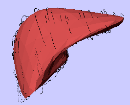 |
The liver reconstructed as a 3D object using piecewise smooth subdivision reconstruction method. The liver was scanned using multiplane abdominal ultrasound techniques in several normal subjects before and after a high-calorie liquid meal10. Liver borders were traced and reconstructed as described for heart borders. Black line segments indicate residual distance between certain "outlier" traced points and the finished smooth surface. |
Back to Top