|
|
|
3D Reconstruction
The Control Mesh - The Platonic Ideal Ventricle
Piecewise Smooth Subdivision Method of Reconstruction
The Control Mesh - The Platonic Ideal Ventricle
To reconstruct the left ventricular endocardium and epicardium, a piecewise smooth subdivision surface is fit to 3D points sampled from the traced borders. This method was developed to avoid the inherent limitations of other methods that restricted their anatomical accuracy or application to different 3D image acquisition techniques. The piecewise smooth subdivision method has been shown to accurately measure volume, and is the only method shown to reproduce LV shape with anatomic accuracy.
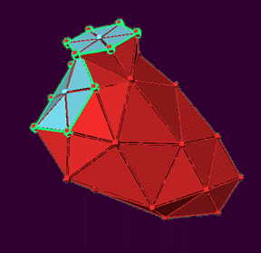
Triangulated control meshes are designed as models for the LV endocardium and epicardium (see below). Each control mesh is recursively subdivided to produce a mesh of 576 faces. The method allows parts of the control mesh to be marked as sharp, allowing for abrupt edges at the junctions between valve planes and the endocardium, and a true vertex at the apex in the final piecewise smooth subdivision surface (Figure 2).
 |
|
The control mesh for the left ventricle endocardium. It serves as the starting point for the piecewise smooth subdivision method. The size, shape, and plane of the triangle surfaces are subsequently modified and refined to approximate the resulting surface described by the 3D traced data points. |
 |
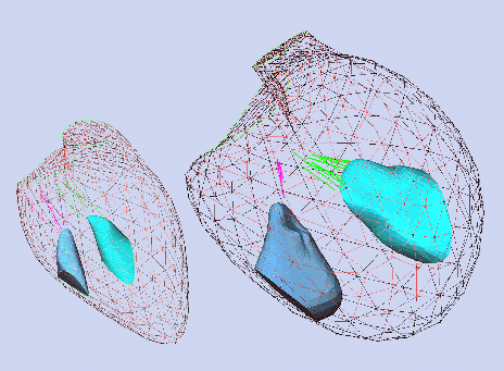
| Wireframe reconstruction of the LV endocardium in a normal subject (left) and a patient with dilated cardiomyopathy (right). Both ventricles are shown to scale. Note the reconstruction of papillary muscles possible with the piecewise smooth subdivision reconstruction method. |
The Piecewise Smooth Subdivision Reconstruction Method
This method makes no assumptions concerning the orientation of the imaging planes, accepting borders traced from rotated, angulated, parallel or randomly oriented images. Thus it is flexible enough to be applicable to any tomographic imaging modality:
Ultrasound and echocardiography
Serial-section transmission light or electronic microscopy
Discrete multiplane confocal microscopy
Computed tomography and magnetic resonance imaging
Nuclear imaging techniques utilizing a high number of acquisition planes
Furthermore, intersecting planes can be analyzed; this is important to verify borders that are better seen in one view than in another. Because we begin with an abstract mesh which embodies knowledge concerning the expected shape of the organ being reconstructed, the method can be used to reconstruct a variety of solid elements:
Heart chambers, vessels, valves, and structures within these
Hollow or solid organs such as the stomach and liver
Other discrete soft tissue structures; e.g. the gastrocnemius muscle in the leg.
The piecewise smooth subdivision reconstruction method allows triangular facets on the reconstructed surface to be labeled to reflect their location on or near anatomic landmarks. This allows analysis of subregions of an organ, and comparison of the same regions of the organ between serial studies or between different subjects. This can best be seen as applied in regional LV shape analysis by Aikawa et al.3
The mitral valve annulus is reconstructed as a smooth curve using four-harmonic Fourier series approximations in each of the three spatial coordinates x, y, and z. The independent variable in each Fourier series is the path length along a polygonal line connecting the actual traced data points. The same algorithm is used for the tricuspid annulus, which also has an irregular, nonplanar ring shape. Points traced from the aortic and pulmonic valve annuli are fitted to a circle.
 |
|
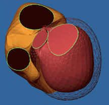
3D reconstruction of the RV (gold) shown with LV endocardium (red solid) and epicardium (pink wireframe). The three structures were traced simultaneously with other cardiac structures; users may reconstruct any one or more subsets of the total traced data for a given patient study. |
Back to Top
|


