Faculty M–Z
The Graduate Program In Neuroscience is comprised of an interdisciplinary faculty from a large variety of departments and 5 partner institutions. If you are interested in joining the program as faculty, please see the requirements and application process.
* Faculty member is currently accepting students in their lab for the 2024-2025 academic year.
i Denotes faculty has funding that is able to support international students.
Michael Manookin | Neural circuits, cells, and synapses that mediate early visual processing.

Research:
Neural computations, circuits, and synaptic physiology of the retina. My lab is focused on understanding how parallel neural circuits in the retina contribute to our visual perception of color, form, and motion. We are currently studying the retinal circuits that contribute to tri-chromatic color vision and motion vision.
Ludo Max | The role of sensorimotor integration in human motor learning and motor control, with an emphasis on auditory-motor learning in speech production and visuo-motor integration in limb movements.

Research:
Research projects conducted in the University of Washington’s Laboratory for Speech Physiology and Motor Control (Max Lab), or through collaborations between this and other laboratories, focus on the neural and sensorimotor processes underlying the control of orofacial and laryngeal movements involved in speech production as well as on human voluntary movements in general. The two major research programs that form the main focus of the laboratory are designed to examine (a) the sensorimotor control and organization of the multiple articulatory and phonatory actions contributing to normal speech production, and (b) the neuromotor and neurophysiological mechanisms underlying stuttering. The work on stuttering also provides additional opportunities to study the neural processes involved in motor control, for example through studies of the effects of dopamine-related pharmacological agents on speech fluency and on both speech and nonspeech motor control. Experimental questions are addressed through the combined use of a variety of available analysis procedures and techniques such as, for example, kinematic and electromyographic analyses of orofacial and limb movements, mechanical perturbations of such movements, electroencephalographic recordings of cortical brain activity, and acoustic analyses of the speech output. Examples of currently ongoing projects include analyses of speech and nonspeech sensorimotor adaptation in normal motor control (e.g., using real-time perturbations of auditory and/or proprioceptive feedback during speech production) and speech and nonspeech movement analyses in stuttering versus nonstuttering adults.
G. Stanley McKnight | The role of intracellular signaling systems in the neuronal circuits that affect feeding and energy metabolism.

Research:
The McKnight lab studies neuronal signal transduction pathways that are regulated by the cAMP/PKA system. One project focuses on the mechanisms that regulate feeding and energy balance in mice using molecular genetic approaches. The cAMP/PKA pathway modulates the sensitivity of neurons in the hypothalamus to leptin and this results in a lean phenotype and resistance to diet-induced obesity. We are trying to define the underlying mechanisms that affect leptin-modulated signal transduction and gene expression. A second project involves the role of PKA and PKA anchoring proteins (AKAPs) in hippocampal dentate granule (DG) cells and their mossy fiber projections. The DG cells participate in contextual pattern recognition and mice with a disruption of a presynaptic AKAP that localizes PKA to the mossy fibers exhibit pattern separation deficits and altered mossy fiber LTP. Changes in gene expression in neurons involved in feeding, energy expenditure, or memory and learning are being monitored using a ribosome-tagging strategy (RiboTag) that we developed.
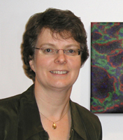
*Kathleen Millen | The Millen laboratory uses molecular genetic approaches to explore the pathogenesis of congenital birth defects of the human and mouse brain and to study genes essential for normal neurodevelopment.
Research:
We are interested in the genetic and developmental basis of structural birth defects of the brain both in humans and mice. We have a specific interest in malformations of the cerebellum, a brain structure that lies between the brainstem and the cerebrum and plays important roles in sensory perception, motor output, balance and posture in addition to cognition and emotion. The relative anatomic simplicity of the cerebellum makes this important brain structure an ideal system to study neural development. Further, the mechanisms that drive cerebellar development are shared by more complex regions of the brain, including the cerebral cortex. Together with Dr. William Dobyns, we maintain the world’s largest clinical database of human cerebellar malformation patients. These birth defects cause neurodevelopmental delays and contribute to Autism. Our resource has enabled us to find the genes that cause these relatively common, yet poorly understood birth defects. In parallel we are also studying mutant mice with cerebellar and other brain malformations to 1) understand how brain malformations arise during development and 2) decipher basic developmental mechanisms that regulate normal brain development. One of our major findings from these studies is that posterior skull development and cerebellar development are fundamentally interdependent. Signals from the developing skull are essential to regulate development of the adjacent cerebellum. Ongoing experiments in the lab are aimed at identifying these signals and their downstream molecular pathways as one means of finding additional human disease causative genes. Finally, Dr. Millen’s group is working on mouse embryonic stem (ES) cell technology to more efficiently and rapidly generate mouse models of human genetic disease. By combining the power and strengths of both mouse and human genetics, our studies are leading to a more comprehensive understanding of the basic biology and genetics of neurodevelopmental disabilities.
*Dana Miller | We use C. elegans to understand how changes in environmental conditions are integrated into organism physiology, and how these response modulate cellular processes involved in neurodegeneration.

Research:
Organisms monitor external conditions and occasionally must initiate adaptation, often at the cellular level, to adapt to new environments. The Miller lab works to identify neuronal mechanisms that coordinate cellular adaptation to changing conditions. Our goal is to understand the pathways that can maintain neuronal and organismal homeostasis in stressful conditions. We currently focus on mechanisms that coordinate adaptation to low oxygen (hypoxia), and the mechanistic basis for the protective effects of hydrogen sulfide signaling. Neurons are particularly sensitive to damage from hypoxia, which contributes to cellular damage and death resulting from stroke. We have also recently become interested in elucidating how these pathways influence protein aggregation and neurodegeneration. We primarily use the nematode C. elegans as a model to discover novel factors that are involved in these processes. The worm is a facile system to map out neuronal pathways that coordinate organism adaptations to hypoxia, as well as the factors that mediate the fundamental cellular processes that contribute to neurodegeneration. We are also mapping the neuromodulators that coordinate organism-wide adaptive responses to adverse hypoxic conditions.
Sheri J.Y. Mizumori | Syst, Cog, M-E-S

Research:
The general goal of this lab is to understand neurobiological mechanisms of plasticity as it relates to learning and memory. We have been studying spatial navigation by rats as a model behavior for understanding how multiple neural systems contribute to complex learning. Adaptive navigation involves the coordination of many processes. Our studies have examined 1) the nature of the sensory input during navigation (e.g. Mizumori & Williams, 1993; Cooper et al., 1998), 2) the integration of current sensory information with past knowledge about an environment (e.g. see Mizumori et al., 1999a,b), and 3) the behavioral implementation of highly processed spatial information (e.g. Mizumori et al., 1999, in press). More recently, we have been considering these processes in terms of the flexibility of underlying neural representational systems during shifts in cognitive strategy or task demands. Other studies have evaluated spatial performance and properties of neural representational systems of aged and brain-damaged animals (e.g. Mizumori et al., 1995, 1996). The majority of our experiments involve recording single unit extracellular activity during behavioral performance.
Cecilia Moens | Developmental genetics of brain patterning in the zebrafish.
Research:
The vertebrate brain contains neuronal representations of the outside world, known as topographic maps, in which the positions of neurons in a projecting field corresponds to the positions of their projections in the target field. These maps form during development, typically through the use of spatial cues that guide axons in a point-to-point matching process. We have discovered a novel “temporal matching” mechanism of topographic mapping that guides cranial motor neurons of the vagus nerve to their target muscles in the pharyngeal arches through the coordinated timing of guidance cue expression in the target field and corresponding receptor expression in the motor neurons. We are studying the genetic, cellular and activity-based mechanisms underlying the formation, refinement and regeneration of vagal reflex circuits as well as other axon guidance events. For these studies we use the transparent zebrafish embryo, which is exquisitely accessible both to genetic manipulation and to high-resolution live imaging of single neurons, their growing axons, and their activity in response to spatially localized stimulation.
*Claudia Moreno | C-M

Assistant Professor
Department of Physiology & Biophysics ✉
Preferred Pronouns: She/Her/Hers
Molecular mechanisms of aging.
Research:
The goal of Dr. Moreno lab is to understand how the function and regulation of ion channels change during the natural process of aging. Aging comes with a vast set of impairments, hearing loss, cardiac dysfunction, and hypertension, are only a few on the list. Most of these impairments are caused by a loss on the capacity of excitable cells to generate and/or propagate electrical signals. As ion channels are the basis of electricity in our body, Dr. Moreno’s team studies how aging affects ion channel function and how these changes can lead to the onset of aging-related pathologies.
To understand the link between ion channel function and the aging process, Dr. Moreno’s team studies one of the most electrical active tissues in the body, the pacemaker of the heart. On average, the human heart beats 100.000 times a day and 2.5 billion times during an average lifetime. Each heartbeat is initiated in the heart’s pacemaker, a few thousands of pacemaker cells that drive the contraction of the more than 2 billion cardiomyocytes of the heart. Aging leads to a decrease in pacemaker activity, and in pathological cases to pacemaker dysfunction, which accounts for more than 60% of the implantations of artificial pacemakers worldwide. Dr. Moreno’s team combines electrophysiology with super-resolution imaging to study age-related changes in the electrical function of the pacemaker and its regulation by the nervous system.
Chet Moritz | We are developing neuroprosthetic technology for the treatment of paralysis and other movement disorders.

Research:
Motor paralysis from stroke or spinal cord injury can be severe and long-lasting, despite damage to a relatively small area of the nervous system. Our goal is to develop neuroprosthetic devices capable of bypassing these damaged areas and restoring volitional control of movement to paralyzed limbs. We have recently demonstrated that this approach is feasible by using activity recorded from motor cortex to directly control electrical stimulation of paralyzed muscles. In addition to replacing lost motor function, we are also attempting to guide and promote the regeneration of damaged neural tissue. Targeted electrical microstimulation can be used to increase the strength of synaptic connections among neurons via mechanisms of Hebbian plasitcity. We are investigating whether this synchronous stimulation, applied across an injury site, can guide neurons to make connections with appropriate targets. We are also testing novel methods for the physical therapy and rehabilitation of movement disorders. We have developed a portable visual feedback device to train children with cerebral palsy (CP) to produce functional muscle synergies. By connecting the activity of impaired muscles to control the movements of popular computer games, we are able to improve volitional control of coordinated muscle activity.
*i Mahmud Mossa-Basha | Neurovascular imaging and its impact on patient outcomes.

Research:
My research and my lab’s research is primarily focused on neurovascular imaging. We specifically work in advanced neurovascular imaging applications, including vessel wall MR, imaging post-processing and deep learning software development for improving patient outcomes, specifically in stroke and cognitive impairment. Our work covers intracranial vasculopathies and extracranial carotid disease. Post-processing algorithms we have developed facilitate automated and semi-automated quantitative image review, enhance and accelerate image review, and aid in vasculopathy diagnosis. We also work to utilize tools developed in the lab and validate their application in clinical datasets to better elucidate disease pathophysiology, longitudinal evolution of vasculopathies, and implications in future events. Our lab looks to recruit talented students to bolster our excellent team. We are looking for students well versed in both technical development, health services research, and clinical investigation of neurovascular disease to enhance the many facets of research in our lab.
Gabe J. Murphy | We determine how particular synapses, cells, and circuits organize and extract the information that enables visually-guided behavior.

Affiliate Assistant Professor
Department of Physiology & Biophysics ✉
Preferred Pronouns: He/Him/His
Neuroscience Focus Areas:
Excitable Membranes and Synaptic Transmission, Neural Circuits, Sensory Systems
Research:
We characterize the selectivity with which cortical and subcortical neurons respond to visual stimuli and the degree to which those responses vary as a function of behavioral state and/or task. Parallel efforts characterize the physiological properties that underlie neuronal response properties – i.e., neurons’ intrinsic biophysical characteristics, the probability and specificity with which they are connected to one another, and the strength and dynamics of signaling between synaptically-coupled neurons. This insight, and assaying the effects of manipulating signals within and/or between neurons, enables us to both form and test hypotheses about how the structure of the nervous system gives rise to its function.
*Gregory J. Morton | Neural mechanisms regulating energy balance and glucose metabolism.
Research:
The Morton lab studies the role of the brain in the control of energy- and glucose homeostasis and how defects in these control system contributes to the development of obesity and diabetes. The overarching hypothesis is that the central nervous system (CNS) senses and receives afferent input from hormonal and nutrient-related signals that convey information regarding both short-term and long-term energy availability and energy stores. In response to this input, the brain engages neuroendocrine, autonomic and behavioral responses that regulate energy intake, energy expenditure, glucose production and glucose uptake in order to maintain energy balance and glycemic control. Our current research: 1) identifies the CNS mechanisms and neurocircuits that mediate these responses; 2) examines how this is integrated with other homeostatic systems such as thermoregulation and circadian rhythms and 3) determines how the brain communicates this information to peripheral tissues. To accomplish this, we utilize state-of-the-art neuroscience approaches, including both “optogenetics and DREADD” methodologies to selectively activate and inhibit specific neuronal populations in combination with genetic, molecular, immunohistochemical and imaging (i.e., fiber photometry) techniques and comprehensive energy balance and glucose-metabolic phenotyping.
*Jay Neitz | Sens & Per

Research:
We are interested in the genetic basis of normal vision and vision disorders. In our laboratory, the latest technology is brought together from molecular genetic, biochemical, imaging, electrophysiological and behavioral approaches. We are using cutting-edge molecular genetics to discover genes that underlie vision loss and gene targeting in mice to dissect the cause of those disorders. Recognizing that the function of the nervous system ultimately involves interplay between genes and the environment, lab members are also seeking potential avenues in which the visual system can respond to environmental influences to restore or even expand neural function. Team members are evaluating the effectiveness of special lenses and filters for preventing vision loss in human patients. In other experiments, we are developing gene therapies with the eye as a model target organ and we are currently at the forefront in research using gene replacement therapy targeting cone photoreceptors in primates. We are optimistic about the potential of gene therapy both as a powerful tool in research and ultimately as a treatment for both stationary and neurodegenerative disorders of the visual system.
Maureen Neitz | Biology of vision and vision disorders.

Research:
We are interested in the genetic basis of normal vision and vision disorders. In our laboratory, the latest technology is brought together from molecular genetic, biochemical, imaging, electrophysiological and behavioral approaches. We are using cutting-edge molecular genetics to discover genes that underlie vision loss and gene targeting in mice to dissect the cause of those disorders. Recognizing that the function of the nervous system ultimately involves interplay between genes and the environment, lab members are also seeking potential avenues in which the visual system can respond to environmental influences to restore or even expand neural function. Team members are evaluating the effectiveness of special lenses and filters for preventing vision loss in human patients. In other experiments, we are developing gene therapies with the eye as a model target organ and we are currently at the forefront in research using gene replacement therapy targeting cone photoreceptors in primates. We are optimistic about the potential of gene therapy both as a powerful tool in research and ultimately as a treatment for both stationary and neurodegenerative disorders of the visual system.
*John Neumaier | Dis, M-E-S

Research:
My laboratory is studying the regulation of serotonin receptors in rat brains in animal models of psychiatric illnesses. We use techniques that span molecular to behavioral levels of analysis. Our strategy is to explore the reciprocal relationship between receptor expression and behavior using techniques that focus on discrete brain regions. There are currently three main projects in the laboratory:
- The role of 5-HT1B autoreceptors in stress-related behaviors associated with stress and depression. We use viral-mediated gene transfer to manipulate 5-HT1B expression in clusters of serotonergic neurons that project to different brain regions and determine the behavioral and physiological outcomes. We have been focusing recently on how these autoreceptors regulate serotonin transporter function using electrochemical and biochemical assays. We are now adapting these methods to transgenic mice that conditionally express 5 HT1B receptors.
- The role of 5-HT1B and 5-HT6 receptors in drug reward mechanisms. Nucleus accumbens and dorsal striatum neurons express these receptors heavily and manipulating their expression with targeted gene transfer alters the rewarding properties of cocaine, amphetamine, and alcohol. We are manipulating these receptors using RNAi (knockdown) or overexpression constructs in pathway-specific viral vectors and measuring the resulting changes in addictive-like behavior using operant conditioning, cocaine self-administration, and others.
- Novel receptors manipulate brain function. We are using light-activated receptors and RASSLs in combination with pathway-specific viral vectors to study how specific groups of neurons participate in complex emotional behaviors relating to stress and addiction.
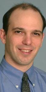
Jeffrey Ojemann | Electrocorticography studies of cognition and brain-computer interface.
Research:
Our lab uses electrocorticography (ECoG) to answer basic neuroscience questions as well as to develop tools for clinical and rehabilitative applications. ECoG, which is used for long-term clinical monitoring of epilepsy patients, provides a unique opportunity to collect intracranial cortical data during awake human behavior in both adults and children (as young as age 2). The group includes researchers from a wide range of backgrounds including neurosurgery, neurology, rehabilitative medicine, engineering, neuroscience, and physics. A major focus of the group is brain-computer interfaces; current projects include learning mechanisms, tactile feedback, and recursive stimulation. Also under investigation are more fundamental questions about the cortical representation of simple and complex hand movements, the dynamics of cognition, language, and higher-order nonlinear interactions between brain areas, and how these phenomenon change with age. Other projects include the integration of ECoG and fMRI and studies of temporal lobe epilepsy.
Jaime Olavarría | Auditory brain sciences and neuroengineering Structure, function, and development of topographically organized circuits in the mammalian visual system.

Research:
In my laboratory, we study the organization, function, and development of neuronal pathways in the mammalian central visual system. Our recent work in primates has employed anatomical and physiological techniques to investigate to what extent visual pathways subserving different functions are segregated, or intermixed, at cortical and subcortical processing stages. We are also interested in studying the role of activity cues on the development of organized cortico-cortical projections in the visual cortex. We are investigating the hypothesis that the neonatal specification of patterns of interhemispheric connections through the corpus callosum depends upon interhemispheric correlated activity driven by visual input. Our efforts include identifying the role of spontaneous and visually evoked retinal activity in the establishment of retinotopically organized patterns of callosal linkages, as well as the cellular mechanisms underlying the effects of activity cues on the development of cortical connections.
Shawn Olsen | Cortical mechanisms of visual behavior and cognition.

Research:
Behavior and cognition are the results of dynamic neural activity in the neocortex and interconnected subcortical structures. We use the mouse visual system as a model to understand the mechanistic basis of these processes. We train mice to perform visual behaviors including stimulus detection and discrimination, and we are actively developing behavioral paradigms to study cognitive processes such as object recognition, spatial attention, and learning. In the context of visual behavioral tasks, we monitor neural activity using both widefield fluorescence imaging and 2-photon calcium imaging. Finally, we use optogenetics to manipulate the underlying circuitry to examine causal relationships between behavior and specific cell types and circuits. We are particularly interested in higher-order cortical visual areas and the role of feedback and interareal projections between cortical areas.
*i Amy Orsborn | Syst, Comp, Motor

Research:
The laboratory works at the intersection of engineering and neuroscience to develop therapeutic neural interfaces. The lab explores neural interfaces as adaptive closed-loop systems that engage neural plasticity and adaptation. We use engineering approaches to leverage neural adaptation for system performance and use neural interfaces as a tool to study neural mechanisms of learning at the systems level. We primarily focus on the sensorimotor system, sensorimotor learning, and interfaces to restore sensorimotor function. The lab also specializes in system integration for advancing neurotechnologies to study neural circuits in awake primates for basic science and human translation. We use state-of-the-art techniques to study neural circuits during behavior in primates, including optogenetics, integrated multi-scale and multi-modal neural measurements/manipulations, large-scale recordings, wireless in-cage recordings, high-dimensional motion tracking, and closed-loop adaptive interfaces.
Richard Palmiter | Our laboratory uses mouse genetic models and viral gene transfer to dissect neural circuits involved in innate behaviors.

Research:
Our laboratory uses mouse genetic models and viral gene transfer to dissect neural circuits involved in innate behaviors. We start by making genetically engineered mice that target the expression of Cre recombinase to genes that are expressed in specific subsets of neurons, typically genes encoding neuropeptides or their receptors. We then stereotaxically inject viruses expressing Cre-dependent genes (e.g., genes encoding fluorescent proteins, genes that allow activation or inhibition of neuron activity by light or chemicals, or genes that prevent all neurotransmission or kill neurons) into brain regions of interest. The aims of these studies are to (a) visualize where the neurons are located and where they project their axons, (b) record the activity of neurons in real-time based on calcium-induced fluorescence, and (c) evaluate the behavioral/physiological consequences of activating or inhibiting those neurons either transiently or permanently. We use combinations of these techniques to delineate neuronal circuits that control specific behaviors. For example, selective stimulation of neurons that express the agouti-related protein (AgRP) promotes feeding, whereas stimulating a different population of neurons that express calcitonin gene-related peptide (CGRP) inhibits feeding. The CGRP neurons that reside in the parabrachial nucleus mediate virtually every threat that we have examined, including real threats (pain, itch, food poisoning) to potential threats (novel food, or cues that have been associated with pain). These CGRP neurons have been shown to mediate the unconditioned stimulus in classical taste- and fear-conditioning experiments. Consequently, they are important for generating taste and fear memories. Current experiments are directed toward identifying the relevant downstream targets of CGRP neurons and discerning how they are involved in mounting appropriate responses to various threats. We are also interested in the functions of other neurons that reside in the parabrachial nucleus that transmit taste, temperature, salt, and water balance signals to the forebrain.
*Jay Parrish | We are broadly interested in understanding the form and function of somatosensory neurons in Drosophila.
Research:
We are broadly interested in understanding the form and function of somatosensory neurons in Drosophila, focusing on the following topics:
- Size control in Dendrites
- Compartmentalization of Dendrite Growth and Patterning
- Substrate control of Dendrite growth
- Dendrite-Substrate Interactions
- Diversity of somatosensory neurons
*i Anitha Pasupathy | Syst, Sens & Per

Research:
Humans gather most information about the world through their eyes. Our brains effortlessly, and rapidly, make sense of the patterns of light that enter our eyes. While this task seems natural to us, it is an amazing feat of computation that no engineer or modern computer has yet been able to approach. How and why is the brain so efficient at understanding the visual world? To answer this question, research in my laboratory focuses on the neural basis of visual shape perception and recognition: the crucial ability to identify and recognize objects from all angles, distances, and in almost any lighting condition. We use single cell neurophysiological studies in awake monkeys, behavioral manipulations, computational modeling and reversible inactivation techniques to investigate how the information reaching our eyes is represented in the neural activity patterns in the brain, how these representations are transformed in successive stages and finally how these representations inform behavior. In addition to shedding light on how visual shape information is processed, these experiments are providing insights into the overall computational capabilities of the primate brain.
David Perkel | Syst, Motor

Research:
I am interested in the detailed cellular mechanisms by which brains learn things. We are using vocal learning in songbirds as a model system for vocal learning in humans, and also for motor learning in general. Young songbirds learn their song first by memorizing the song of a nearby individual, usually the father. Later, they begin to vocalize and slowly match their own vocalizations to the memory of their “tutor”. The tutor does not need to be present during the practice phase, but the bird needs to have intact hearing. When the young bird achieves a good match of the tutor song, his song becomes highly stereotyped, and its maintenance becomes somewhat less dependent on hearing. Extensive information concerning the brain structures involved in song production and learning, combined with detailed subcellular understanding of synaptic plasticity phenomena such as long- term potentiation (LTP) in the mammalian hippocampus, allow us to make testable hypotheses regarding the cellular interactions that underlie this behavior. We are using in vitro brain slices obtained from zebra finches to study synaptic mechanisms and plasticity in this system. The goal of this approach is to link cellular and synaptic events with behavior, and we plan to use knowledge gained from in vitro work to guide experiments to investigate the role of synaptic plasticity in song learning in vivo.
A second project in the lab concerns a circuit essential for vocal learning but not adult song production, the so-called anterior forebrain circuit. We are testing the hypothesis that this pathway corresponds to the mammalian cortico-basal ganglia-thalamocortical loop. We have provided strong evidence for functional similarities in the neurotransmitters used in some portions of this pathway and are continuing to explore the implications of this hypothesis at cellular, systems and behavioral levels. Tying this learning circuit to a well-studied pathway in mammals will allow work in avian and mammalian systems to be mutually beneficial.
*i Steve I. Perlmutter | Syst, Dis, Motor

Research Associate Professor
Department of Physiology & Biophysics ✉
Preferred Pronouns:
Understanding and manipulating neural plasticity in mammalian motor systems to develop new therapies that improve recovery after spinal cord injury and brain damage.
Research:
Motor deficits severely impact the quality of life of people with damage to the brain and spinal cord, yet current treatments produce limited improvements in movement abilities. Although the body’s natural recovery processes after injury do not cause spared neural pathways to achieve their fullest potential for restoring function, substantial behavioral gains can be achieved with small but opportune changes in cortical and spinal organization. We are investigating strategies for inducing plasticity in normal and lesioned motor systems using activity-dependent, targeted, electrical and optical stimulation and delivery of neuromodulators and neurotrophins. Our goal is to develop neuroprosthetic therapies that exploit the nervous system’s capacity for plastic rewiring to improve motor function after stroke, traumatic brain and spinal cord injury.
In addition, we are interested in interdisciplinary, collaborative approaches to facilitate neural regeneration in corticospinal pathways after spinal cord injury. We are currently using neural cell cultures to understand activity-dependent mechanisms of neurite growth and synaptogenesis.
Our current work on neural plasticity builds on our experience studying the neural mechanisms of voluntary hand and arm movements. Primates generate an incredibly varied repertoire of motor behaviors. We have elucidated principles of cortical and spinal information processing that accomplish flexible, coordinated motor control in non-human primates.
Our lab uses neurophysiological, behavioral, anatomical, computational, and genetic techniques in studies in rodents and non-human primates. We have active collaborations with cell and gene biologists, neurosurgeons, and engineers designing devices for brain-computer interfaces.
*i Paul Phillips | Syst, M-E-S

Research:
The key focus of our lab is to precisely define the role of dopamine neurotransmission in motivated behaviors and decision making, and to use this information to address how physiological processes that control this transmitter may alter behavior. A major component of our research is the study of rapid (phasic) dopamine transmission during addictive behaviors. One of the main tools we use is fast-scan cyclic voltammetry. This is a rapid electrochemical technique that can detect dopamine several times a second and chemically resolve it from other electroactive species. When used in awake rodents, this technique has been particularly useful for elucidating the precise temporal relationship between released dopamine and behavioral events. Since the temporal resolution is sufficient to capture chemical information on the physiological time scale, it is also well suited for studying the dynamics of the system and probing short-term presynaptic plasticity.
Chantel Prat | My research investigates the biological basis of individual differences in language and cognitive abilities.

Research:
Human thought is characterized by its flexible, dynamic nature. My research at the Cognition and Cortical Dynamics Laboratory (CCDL) attempts to understand how the brain learns and adapts to deal with the ever-present fluctuations in the environment. I am particularly interested in individual differences in language and cognitive capabilities, and how they are reflected by differences in brain functioning. In addition, my current research investigates the overlap between language and general information processing abilities by exploring improvements in general executive functions in individuals who develop bilingually, and deficits in general executive functions in language-impaired populations such as autism spectrum disorder. The CCDL utilizes multiple methods and approaches including functional magnetic resonance imaging (fMRI), transcranial magnetic stimulation (TMS), and individual differences research to collect converging evidence about the biological nature of human thought.
*iMarco Pravetoni | Therapeutics for substance use disorders, overdose, and other unmet public health threats.

Research:
My NIH-funded research program in Medication Development for Substance Use Disorders (SUD) focuses on these major efforts: 1) development and translation of vaccines, monoclonal antibodies (mAb), and small molecules to treat or prevent SUD, opioid use disorders (OUD) and overdose, 2) mechanisms and biomarkers underlying or predicting efficacy of immunotherapeutics and medications in pre-clinical models and OUD patients, and 3) novel strategies to enhance vaccine or medication efficacy, including immunomodulators, small molecules, adjuvants, nanoparticles, polymers and other delivery platforms. In the SUD field, I am also involved in team efforts to develop point of care devices (e.g., laminar flow or microfluidic assays) to detect drugs of abuse for diagnostics, forensic, or other field applications. These approaches are applied to other targets such as chemical threats, cancer, SARS-CoV-2 and other infectious agents.
Daniel Promislow | We use quantitative genetic and systems biology to identify naturally occurring modifiers of neurodegenerative disease in the fruit fly.

Research:
I am an evolutionary biologist interested in the role of natural genetic variation in patterns of aging. The study of aging has led our lab in many directions, including the analysis of behavior and brain function. Two specific Drosophila projects focus on issues related to the brain and behavior. Over the past 25 years, researchers have identified insulin signaling as a key pathway influencing patterns of aging in model organisms. Recent studies have found that insulin signaling also appears to play an important role in the development of traits that are important in mate choice. We are using the fruit fly as a model system to explore the role that insulin signaling plays in shaping mate attractiveness and mate preference in natural populations. These studies include the use of molecular and evolutionary genetic approaches to better understand the connection between insulin signaling, sensory perception, chemical communication, mate choice, and fitness. This study also takes advantage of metabolomic profiling as a way to link genotype to phenotype. The second set of studies, in collaboration with Leo Pallanck in the Department of Genome Sciences at UW, uses statistical network analysis of metabolomic profiles to better understand the mechanistic pathways associated with neurogenerative diseases in flies. Researchers have identified numerous mutants in the fly that recapitulate neurodegenerative diseases seen in humans. We are using metabolomic approaches to identify natural variants associated with neurodegeneration and to better understand the functional mechanisms by which these mutations give rise to neurodegeneration.
*i David Raible | Zebrafish mechanosensory hair cell development, damage and regeneration: models for hearing loss.

Professor
Department of Biological Structure ✉
Preferred Pronouns:
Neuroscience Focus Areas:
Cell and Molecular Neuroscience, Developmental Neuroscience, Sensory System
Research:
Hair cells of the inner transduce mechanical stimuli to electrical signals transmitted to the brain. Hair cells of the auditory organs respond to sound stimuli for hearing perception; those in the vestibular organs respond to gravity and head movements for the perception of balance. Hair cells are so named because they have actin-rich protrusions, stereocilia, at their apical end. Displacement of stereocilia opens ion channels resulting in depolarization and release of transmitter from synapses at the basal end of the cell to terminals of innervating afferent nerves. Damage and loss of hair cells are leading causes of hearing and balance disorders, which affect over 40 million people in the US. Hair cells are susceptible to environmental insults, including noise, chemical exposure, and accumulated damage during aging. Genetic disorders are common, affecting more than 1:500 children. Hair cell loss in humans is irreversible.
We use the zebrafish lateral line system to study hair cells. Like those of the inner ear, lateral line hair cells respond to fluid movement and synapse with afferent neurons. However, as they are located on the body surface they are readily accessible for visualization and manipulation. In contrast to humans, zebrafish regenerate their hair cells from a dedicated pool of precursors. We use genetic manipulation such as CRISPR and in vivo imaging using encoded fluorescent reporters to study hair cell development, death, and regeneration. Our studies are leading to therapeutic approaches to prevent hair cell damage and potentially restore function.
Akhila Rajan | Fat-brain communication: how fat signals to the brain.

Research:
Taking advantage of the deep, and unexpected, evolutionary conservation between fat-brain communication in flies and humans (Rajan and Perrimon, Cell, 2012), we explore how adipokine (fat hormone) signaling modulates many aspects of physiology. By investigating fat physiology, we have uncovered new insights in the areas of: 1. of cell biology, we discovered a mechanism by which fat cells couple nutrient state to adipokine retention (Rajan et al., Dev Cell, 2017; Poling et al, biorxiv, 2021). 2. and neurobiology by uncovering how two hormones act on the same synapse to regulate its structure and shown that this mechanism regulates body weight (Brent and Rajan., Cell Metabolism, 2020). Now, the lab is actively investigating following questions:
1. How does adipokine nuclear accumulation regulate post-feeding hunger motivation? 2. Modulators and regulators of adipokine transit across the blood-brain barrier. 3. Mechanistic understanding of how a metabolic dysfunction causes cognitive decline. Specifically, we investigate the connection between adipokine transit into the brain and how it might neuroinflammation. To address these questions, we deploy an inter-disciplinary toolkit, both in vivo and using cell culture-based systems. Techniques we use regularly are fruit fly genetics, behavioral assays, quantitative imaging, lipid chromatography, mass spectrometry etc., in conjunction with emerging approaches including super resolution microscopy and cutting-edge methods in genomics and proteomics.
Jan-Marino (Nino) Ramirez | Understanding the neuronal basis of a variety of brain functions to find novel ways to treat and cure neurological disorders in children, including epilepsy, Rett syndrome, brain tumors, and sudden infant death syndrome.

Research:
With the introduction of molecular approaches into the field of neuroscience, an unexpected variety of receptor and ion channel subtypes have been discovered that are developmentally regulated within the central nervous system. The relevance of these findings to the function of neuronal networks is still unclear. In a close combination of studies at the cellular and systems level, our laboratory examines how diversity at the cellular level may lead to ontogenetic changes at the network level.
Over the past decade, we have analyzed cellular mechanisms in neuronal networks that generate rhythmic motor activity in invertebrates and vertebrates. Our current work focuses on the analysis of the in vitro respiratory network in mice. For this purpose, we isolate acutely the respiratory network in a transverse plane of the mouse medulla. This brainstem slice preparation contains the essential medullary structures involved in cardio-respiratory control and even after the in vitro isolation generates rhythmic activity in rats and mice of all developmental stages (up to the age of 25 postnatal days). This approach permits a direct comparison of the neuronal mechanisms of rhythmogenesis in newborn and more mature mammals. Our experiments indicate e.g. that the hypoxic response, fast chloride mediated inhibitory synaptic transmission, calcium channels, and modulatory processes change postnatally within the respiratory network.
In order to analyze the cellular mechanisms in rhythm-generating neural networks, we employ the currently available electrophysiological and immunohistochemical techniques. The great advantage of the brainstem slice preparation is that single neurons can be visualized that are embedded in a functional neuronal network using infrared-Normarski optics in conjunction with upright microscopes. Thus, voltage-gated and synaptic whole-cell currents, properties of single channels as well as second messenger pathways can be investigated in a functional context to characterize changes in the postnatal development of the respiratory network. Our laboratory is particularly interested in understanding developmental alterations of cellular properties involved in the response of the respiratory network to hypoxia. This response elicits a cascade of molecular events which are regulated by endogenously released neuromodulators such as substance P and endorphins and which result in a reconfiguration of the respiratory neuronal network.
In the future, this slice preparation will enable us to further obtain systems and cellular data from functionally identified respiratory neurons. In combination with cell culture and modern molecular techniques (e.g. expression of channel subtypes obtained from respiratory neurons in oocytes), we expect to gain new insights into principle mechanisms of respiratory rhythm generation and the hypoxic response in mammals. Due to the importance of the respiratory system for the survival of any mammal, progress in this field will not only have important scientific but also clinical implications (e.g. understanding the underlying causes of sleep apnea, periodic breathing, Pickwick syndrome, and sudden infant death syndrome “SIDS”).
Rajesh R.N. Rao | Computational neuroscience, machine vision and robotics, and brain-computer interfaces.

Research:
The goal of my research is to discover the computational principles underlying the brain’s remarkable ability to learn, process, and store information, and to apply this knowledge to the task of building adaptive robotic systems and intelligent machines. Some of the questions that motivate my research include: How does the brain learn efficient representations of novel objects and events occurring in the natural environment? What are the algorithms that allow useful sensorimotor routines and behaviors to be learned? What mechanisms allow the brain to adapt to changing circumstances and remain fault-tolerant and robust? By investigating these questions within a computational and probabilistic framework, one can derive algorithms that provide functional interpretations of neurobiological properties and at the same time, suggest solutions to difficult problems in computer vision, robotics, and artificial intelligence. Some illustrative examples of my research efforts in these directions, along with selected online publications, can be found on my UW Home Page (see link above).
Jeff Rasmussen | C-M, Dev

Research:
The Rasmussen lab uses zebrafish to gain molecular and cellular insights into neuronal plasticity, both during development and following injury. The skin is our largest sensory organ and is densely innervated by somatosensory nerve endings that sense pain and touch. Nerve regeneration is often incomplete following skin injury, and sensory loss is a major complication associated with diabetes and chemotherapy. Using the imaging advantages of the zebrafish system and novel remodeling assays developed in the lab, we have identified interactions between somatosensory nerve endings and several specialized cell types (including osteoblasts, resident macrophages, and mechanosensory cells) within the skin tissue environment. We are currently pursuing the following related projects:
(1) Control of nerve patterning during skin development
(2) Genomic and cellular analysis of nerve regrowth following injury
(3) Development, regeneration, and function of mechanosensory cells
*i Tom Reh | Determination of the mechanisms that control neuronal proliferation and differentiation during neurogenesis of the vertebrate CNS.

Research:
Our lab is focused on the development and repair of the retina, the light-sensitive tissue of the eye. We apply the information we obtain from our developmental studies to develop methods for retinal repair using human embryonic stem cells, induced pluripotent cells, or through the stimulation of endogenous repair mechanisms. We have been particularly interested in the role of the Notch signaling pathway in retinal development and repair. We have developed methods for directing human ES cells to a retinal progenitor fate, and are exploring the molecular mechanisms that further restrict their identity to rod and cone photoreceptors. We have found that the transplantation of photoreceptors derived from human ES cells can restore light response to mice with congenital blindness. Although our studies in amphibians and birds have shown that endogenous repair mechanisms can regenerate retinal cells in non-mammalian vertebrates, recent work from the lab also demonstrates a limited potential for the endogenous repair of the retina after damage, from a population of support cells known as Muller glia. We are working to determine the molecular mechanisms that limit their capacity for regeneration in mammals by determining their differences from non-mammalian vertebrates.
R. Clay Reid | Deciphering how information is encoded and processed in neural networks of the visual system, using behavior, anatomy and physiology.

Research:
Our group studies how the brain processes and makes sense of visual information, using a variety of methods including electrophysiology, imaging, large-scale electron microscopy, and quantitative analysis. Most recently we have used a combination of imaging and anatomical approaches to investigate how the structure of neural connections relates to functional circuitry in the visual cortex.
Fred Rieke | Sen & Per

Research:
The research in my lab focuses on sensory signal processing, particularly in cases where sensory systems perform at or near the limits imposed by physics. Photon counting in the visual system is a beautiful example. At its peak sensitivity, the performance of the visual system is limited largely by the division of light into discrete photons. This observation has several implications for phototransduction and signal processing in the retina:
- rod photoreceptors must transduce single photon absorptions with high fidelity,
- single photon signals in photoreceptors, which are only 0.03 – 0.1 mV, must be reliably transmitted to second-order cells in the retina, and
- absorption of a single photon by a single rod must produce a noticeable change in the pattern of action potentials sent from the eye to the brain.
My approach is to combine quantitative physiological experiments and theory to understand photon counting in terms of basic biophysical mechanisms.
Fortunately, there is more to visual perception than counting photons. The visual system is very adept at operating over a wide range of light intensities (about 12 orders of magnitude). Over most of this range, vision is mediated by cone photoreceptors. Thus adaptation is paramount to cone vision. Again one would like to understand quantitatively how the biophysical mechanisms involved in phototransduction, synaptic transmission, and neural coding contribute to adaptation.
*i Jeff Riffell | Olfactory neurobiology and chemical communication processes.
Research:
Sensory perception of chemical signals strongly influences reproduction, habitat selection, as well as cellular navigation and motility. Indeed, a variety of physiological processes and behaviors are critically dependent on chemosensory signaling mechanisms. Despite the complexity of these processes, the functional principle is the same: detection of chemical stimuli is transduced\ via biochemical signaling cascades and further processed in the brain. The main goal of my lab’s research, therefore, is to understand these signaling mechanisms on a cellular level and at the level of neuronal processing in the brain.
Olfactory processing of complex odor stimuli
An ‘odor’ is composed of a complex mixture of different molecules at various concentrations. In the brain, this information is encoded by spatial (and temporal) activity patterns of distinct neuron populations within the antennal (olfactory) lobe. Our research indicates that the temporal activity of the insect antennal lobe (AL) neurons, similar to that which occurs in the visual system, acts to ‘bind’ the complex mixture representation into a single odor percept. Using multi-electrode extracellular recordings of AL output neurons and behavioral assays, we aim to analyze the neural basis of behavior and neuronal network activity in the AL.
Chemotactic signaling in single cells
At a much smaller scale, we examine chemical communication processes at the level of the single cell where the basic molecular and cellular mechanisms underlying chemotaxis are far less understood. We recently found that members of the odorant receptor (OR) family are key mediators of sperm chemotaxis, and in parallel studies with invertebrates, egg-derived chemical attractants increase sperm-egg encounters and fertilization success. We are currently examining the contribution of ORs in mediating chemotactic behaviors and the molecular basis of this process.
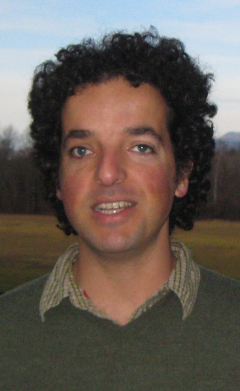
Ariel Rokem | Neuroinformatics, data science, connectomics.
Research:
The brain is a tremendously complex system. In order to understand it we are going to need large amounts of data from many different kinds of measurements. We use data science methods to integrate the information provided by these measurements into a coherent picture. In particular, we develop statistical analysis techniques to decipher the role of networks of brain areas in complex behaviors and in brain disorders. We implement these techniques in robust, efficient, and openly-available computer software.
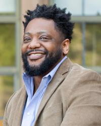
Oliver Rollins | Focuses on ethical, social, and legal issues in the mind and brain sciences, specifically the impacts of race/racism and social difference in neuroscience.
Assistant Professor
Department of American Ethnic Studies ✉
Preferred Pronouns: He/Him/His
Neuroscience Focus Areas:
Neuroethics, Behavioral Neuroscience, Diversity, Equity, and Inclusion (DEI)
Research:
Oliver Rollins is a qualitative sociologist who works on issues of race/racism in and through science and technology. Specifically, his research explores how racial identity, racialized discourses, and systemic practices of social difference influence, engage with and are affected by, the making and use of neuroscientific technologies and knowledges. Rollins’ book, Conviction: The Making and Unmaking of The Violent Brain (Stanford University Press, 2021), traces the development and use of neuroimaging research on anti-social behaviors and crime, with special attention to the limits of this controversial brain model when dealing with aspects of social difference, power, and inequality. Rollins’s current projects focus on 1) the social implications and challenges of operationalizing racial prejudice, implicit bias, and identity as neurobiological processes, and 2) the politics of social justice and (neuro)science, which aims to elucidate the socio-political vulnerabilities and anti-racist promises for contemporary neuroscientific practices. Rollins teaches courses on the social (racialized) implications of science and technology; theories of race and blackness; and neurotics, and biopolitics of biomedical and scientific knowledge.
Jay Rubinstein | Signal processing, physiology, and perception with inner ear implants using both computational modeling and experimental techniques.

Research:
The Rubinstein lab studies cochlear implants and collaborates with the Phillips lab in the study of vestibular implants. The lab uses computational biophysical and empirical modeling, as well as digital signal processing, psychophysics, and speech and music perception studies. Physiological studies are performed in collaboration with the Tremblay lab for cochlear implants in humans and the Phillips lab for vestibular implants in both human and non-human primates. Human subjects for cochlear implant research include both normal hearing listeners, as well as a variety of cochlear implant devices including Hybrid electro-acoustic implants currently undergoing clinical trials at a limited number of sites. The lab has access to the world’s only human subjects implanted with a vestibular neurostimulator. Our goals are to use fundamental neuro engineering principles to improve signal coding in inner ear devices, demonstrate the efficacy of these improvements in humans and translate them to clinical use.
*i Hannele Ruohola-Baker | Regulation of stem cell self renewal and regeneration.
Research:
A critical question in stem cell biology is how stem cells escape cell division and stop signals. We have shown the necessity of the microRNA (miRNA) pathway for proper control of germline stem cell (GSC) division in Drosophila melanogaster. Analysis of GSCs mutant for dicer-1 (dcr-1), the dsRNAseIII essential for miRNA biogenesis, revealed a dramatic reduction in the rate of germline cyst production. These dcr-1 GSCs exhibit normal identity but are defective in cell cycle control. Based on cell cycle markers and genetic interactions, we conclude that dcr-1 GSCs are delayed in the CDK-inhibitor p21/p27/Dacapo-dependent G1 to S transition, suggesting that miRNAs are required for stem cells to bypass the normal G1/S checkpoint. Hence, the miRNA pathway might be part of a mechanism that makes stem cells insensitive to environmental signals that normally stop the cell cycle at the G1/S transition. We are now in the process of analyzing the microRNAs critical for stem cell division and identifying the region of Dacapo 3’UTR responsive to these microRNAs. Germline stem cells reside in a microenvironment, a niche where they undergo asymmetric division to produce the differentiating cells destined to develop into mature eggs. In the absence of injury, it has been thought that these stem cells persist for the life of the organism. We have shown that this is not the case: stem cells are replaced every three to four days throughout the life of the adult fly. The continuous replacement may provide a robust means of maintaining the stem cell population and contribute to its longevity.
Using a genetically tractable Drosophila model for studying muscular dystrophy, we have dissected the function of the Dystroglycan(DG)-Dystrophin(Dys) complex in muscle and in the brain. Genetic and RNAi-based perturbation of DG and Dys causes both cell polarity and muscular dystrophy phenotypes: decreased mobility, shortened lifespan, age-dependent muscle degeneration, and defective photoreceptor path-finding. In the latter case, we find that the DG-Dys complex is essential both in the photoreceptor neurons and the targeting glial cells, suggesting that both cell types are involved in the ECM-based regulation of axon pathfinding. Using a fluorescence polarization assay, we have shown that the DG-Dys interaction is remarkably well evolutionary conserved between flies and humans. Surprisingly structure-function studies of DG revealed that a truncation of the WW-binding motive, thought to interact with Dystrophin is not essential for DG function. However, one mutation to alanine of a proline within an SH3-domain binding site abolishes DG function suggesting a critical interaction with a potential signaling molecule that might regulate the complex. Genetic and biochemical screens are in progress to reveal the critical interacting genes.
Ramkumar Sabesan | Functional imagining of the human retina.

Research:
The mechanisms by which the physical attributes of the world – color, space, motion – are derived from individual cone photoreceptors and facilitated by the postreceptoral circuitry remain unclear. For instance, short-wavelength cones are activated to the same extent by small amounts of blue wavelength light as they are activated by large amounts of red wavelength light. It is remarkable then that the visual system is capable of reconstructing fine spatial detail and rich color experience from the external world even though intensity and wavelength of light are confounded at the scale of single photoreceptors. From a clinical standpoint, photoreceptors are the most vulnerable to blinding retinal diseases. Bringing vision restoration therapies to living humans and their continuous improvement relies on parallel technological innovation in the microscopic visualization and manipulation of retinal cells in vivo.
Our lab uses interdisciplinary approaches to study the functional mechanisms by which photoreceptors and their ensuing visual pathways mediate the most fundamental aspects of vision and how these visual capacities are affected by diseases. To achieve this, we develop and use novel imaging tools that enable the visualization of the structure and function of retinal cells at unprecedented spatial scales. The backbone of the methods pursued by our lab is a technology called adaptive optics – the same tool used by astronomers to peer at small objects in space. Using adaptive optics, we can overcome the optical imperfections that exist in the human eye converting our eyeball essentially into a microscope objective. This gives us the ability to probe living cells in the retina of humans which are about ten times finer than the diameter of a human hair. The image below shows the long, middle, and short-wavelength cones in the retina of a living human obtained using a special optical system equipped with adaptive optics. Ultimately, we aim to use such high-resolution functional assays as sensitive biomarkers for early disease diagnosis, monitoring of disease progression, and efficacy of treatments.
Abigail G. Schindler | Computational medicine, iterative translation, and systems biology to understand traumatic stress and its comorbidities.

Assistant Professor
Department of Psychiatry & Behavioral Sciences ✉
Preferred Pronouns:
Neuroscience Focus Areas:
Behavioral Neuroscience, Cellular and Molecular Neuroscience, Computational Neuroscience, Neurotransmitters, Modulators, Transporters, and Receptors
Research:
We are interested in traumatic stress and its comorbidities (e.g. affective disorders, substance abuse, metabolic dysfunction, neurodegeneration). Using an iterative translation approach, we utilize both human and animal populations and focus on reciprocal connections between the brain and peripheral organs (e.g. liver, gut, lymph) in order to understand adverse outcomes of traumatic stress from a systems biology standpoint. We are especially interested in understanding the role of monoamines (e.g. dopamine, serotonin) and neuromodulators (e.g. opioids, cytokines). We utilize computational medicine approaches (e.g. machine learning, big data, electronic health records, biomarkers) and are committed to open source science.
John Scott | Specificity of synaptic signaling events that are controlled by kinase anchoring proteins.
Research:
My research program focuses on defining the intracellular communication networks that promote specificity in signal transduction events. We have identified a family of A-kinase–anchoring proteins (AKAPs) that target the cAMP-dependent protein kinase (PKA) and other signaling enzymes to specific subcellular sites. AKAPs influence the specificity of second messenger–mediated signal transduction events by targeting enzymes close to their appropriate effectors and substrates. Our lab has made progress in establishing the AKAP model, the functional consequences of PKA anchoring, and the role of AKAP signaling complexes in the coordinate regulation of certain synaptic and cytoskeletal signaling events.
Eric Shea-Brown | Computational and theoretical neuroscience.

Research:
Eric’s group works on the dynamics of neural networks and neural populations. These dynamics are beautiful, and are richly varied from setting to setting – at times governed by mechanisms, we can distill and explain and at times eluding our best analytical tools. Beyond understanding the nonlinear dynamics of neural circuits, we want to understand how they encode and make decisions about the sensory world.
Ongoing projects are on: (1) the time course of decision making in neural networks, seeking both behavioral signatures and neural correlates of decision processing that we can test for in data, (2) collective coding in neural populations, (3) graph-theory tools that link architecture and neural dynamics, with the aim of helping to cut connectomics problems down to size. Making progress on these problems requires a range of perspectives and methods. Eric and his group delight in collaboration with fellow theorists of many different backgrounds, and in close ties with cognitive neuroscientists, biophysicists, and neurophysiologists. These colleagues are developing extraordinary datasets that reveal the brain’s dynamics on a range of scales that would have been unimaginable even a decade ago. As a consequence opportunities for impactful mathematical analysis and modeling currently seem boundless. Eric’s group works within the creative and expansive neuroscience community at UW and collaborates closely with Allen Institute for Brain Science, a unique and inspiring institution that we are lucky to have just across our city.
Andy Shih | Our lab uses imaging approaches to better understand the regulation of brain microvascular health, and the factors that lead to its dysfunction during disease.

Research:
Our research focuses on the small blood vessels that delivery oxygen and nutrients to all reaches of the brain. In the human brain, an estimated 400 miles of blood vessels delivers blood to 100 billion neurons. This immensely complex task can easily go awry during human disease. When we are young, genetic and environmental factors can compromise the normal development of brain vasculature. As we age, leakage and blockage of small vessels can contribute to Alzheimer’s disease and vascular dementia. Our laboratory studies how blood vessels grow, degrade, and respond to injury from birth to senescence. To study the brain vasculature, we use a variety of approaches, including in vivo multiphoton microscopy, optogenetics, 3-D electron microscopy, magnetic resonance imaging, and development of transgenic mouse models. We hope that our research will yield new ways to reduce to improve cerebrovascular function in brain diseases that affect both children and adults.
*Aakanksha Singhvi | Glia in health, aging, and disease

Research:
Glia makes up about half of our brain cells and communicates closely with neurons throughout life, both physically and biochemically. Glia-neuron interactions underlie proper nervous system development and functional maintenance throughout life and disrupted interactions between these two cell types underlie many neurological disorders of development (eg. autism), function (sensory or cognitive impairments), and aging (Alzheimer’s).
Despite this, glial functions in the nervous system and glial-neuron interactions remain poorly understood in molecular detail. Our lab is interested in building a molecular mechanistic framework of how glia and neurons communicate with each other across to regulate sensory perception, neuronal physiology, neural circuit activity, memory formation, and animal behavior.
C. elegans is powerful in vivo model to dissect molecular mechanisms of glia-neuron interactions. In this genetically tractable animal with a mapped connectome, any glia or neuron of choice can be manipulated at single-gene resolution. Effects of such manipulations can be monitored at many levels, from gene networks and –omics, biochemical investigations, cell biology, neural connectivity, in vivo light, super-resolution and functional imaging, EM and sensory animal behaviors, and memory assays. We typically use all of these approaches to understand glia-neuron interactions; and coopt or develop additional methods, tools, and techniques, as needed, to tackle a given scientific question.
Joseph Sisneros | Understanding how the vertebrate auditory system processes species-specific vocalizations and the adaptive mechanisms that are used to optimize the receiver’s sensitivity to social communication signals.
Research:
My research examines steroid-dependent plasticity in the adult auditory system of the plainfin midshipman fish (Porichthys notatus). The vocal midshipman has become a model for identifying the mechanisms of auditory reception, neural encoding, and vocal production shared by all vertebrates. Vocal communication is essential to the reproductive success of the seasonally breeding midshipman, which migrates during the late spring and summer from deep ocean sites into the shallow intertidal zone to spawn in nests under rocky shelters. Males produce long-duration advertisement calls or ‘hums'( to attract gravid females to their nest. The seasonal onset of male advertisement calling in the midshipman fish coincides with a dramatic increase in the range of frequency sensitivity of the female’s sacculus, the main peripheral organ of hearing, thus leading to an increased encoding of the advertisement calls & upper harmonics that can, in turn, improve a female’s ability to detect and locate nesting males in shallow water environments like those where males acoustically court females (Sisneros and Bass 2003; J Neurosci. 23:1049-1058). Approximately one month before spawning, midshipman females undergo ovarian recrudescence and like many other teleost fish show seasonal peaks in circulating plasma levels of both testosterone and 17B-estradiol (Sisneros et al. 2004; Gen. Comp. Endocrinol. 136:101-116).
We now show that non-reproductive females treated with either testosterone or 17R-estradiol exhibit an increase in the sacculus’ frequency sensitivity that mimics the reproductive auditory phenotype (Sisneros et al., in press; Science). A likely site where such steroid-dependent changes in auditory tuning occur is at the level of the hair cell receptor. Thus, the principal goal of my current research is to characterize the frequency response dynamics of midshipman saccular hair cells and determine via behavior experiments how reproductive female midshipmen localize behaviorally-relevant auditory stimuli such as the male’s advertisement (mate) call.
*Stephen Smith | Understanding how the vertebrate auditory system processes species-specific vocalizations and the adaptive mechanisms that are used to optimize the receiver’s sensitivity to social communication signals.

Research:
The SEPS lab studies the molecular biology of the glutamate synapse, with a focus on the molecular mechanisms shared among different genetic causes of autism. The protein products of genes do not act in isolation, but form networks of multi-protein complexes, tiny molecular machines that function onl methods to analyze the behavior of these multi-protein complexes. We use these techniques, along with molecular and genetic approaches, to model dynamic protein interaction networks at the synapse, and to investigate how these networks are disrupted in autism.
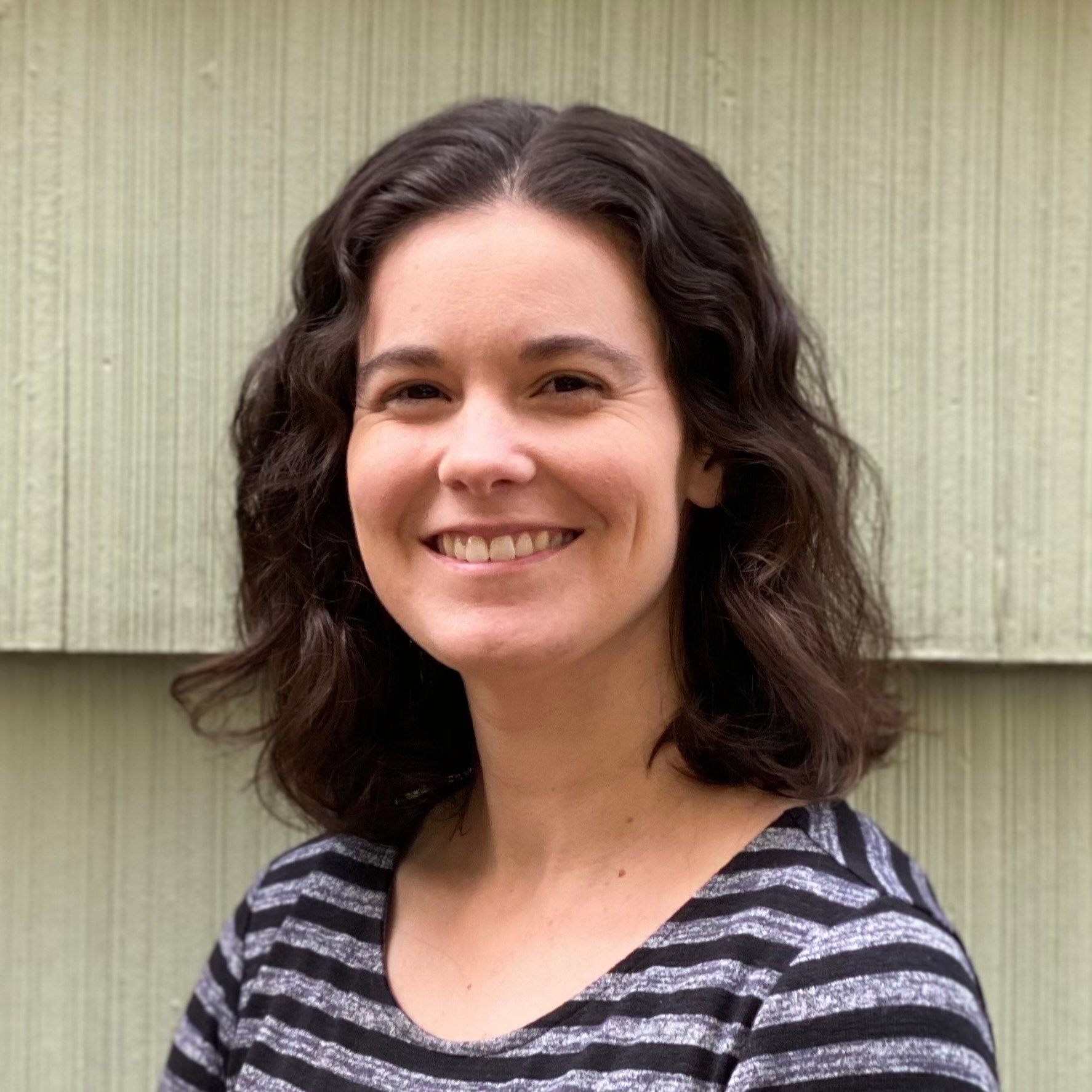
*Marta Soden | Dissecting neural circuits underlying motivated behavior.
Research:
We are interested in better understanding the organization, connectivity, and physiology of neural circuits that regulate motivated behavior. A major focus of the lab is investigating the circuit connections that regulate the activity of midbrain dopamine neurons. We are particularly focused on inputs to the dopamine system that co-release neuropeptides along with fast neurotransmitters, with the goal of understanding how these factors work together to determine circuit output. We also study the function of ion channels that regulate neuronal excitability and responsively to synaptic input. We utilize optogenetics, fiber photometry, transcriptomics, electrophysiology, CRISPR/Cas9 mutagenesis, and other advanced techniques to ask questions about gene function, neuronal excitability, synaptic connectivity and behavior.
William Spain | Transformation of synaptic inputs into patterns of action potential output; information flow within the network of neurons.

Research:
What am I? Where am I? Who am I? Time, Space and Consciousness – the pursuit of science and the folly of art. A reminder lest we lose sight of the “big questions” in our daily labors on form and function. The lab? It deals with both of those issues (form and function). Will the results shed light on consciousness? Doubtful, but let’s keep our minds open for possible connections. After tabling the quest for a complete understanding of consciousness, I have settled to the more pragmatic task of studying the mechanisms by which central neurons code information under normal conditions and how those processes are altered in neurological disease. To that end, my lab is identifying the rules for transducing synaptic input into frequency-coded trains of action potentials in neocortical neurons and brainstem auditoiy relay neurons. Because those neurons perform different functions, their transduction mechanisms contrast sharply. For example, the cortical Betz cells provide the primary motor output to brainstem and spinal cord. Cortical integrative processes converge on Betz cells; thus, they sit in a position critical for the summing of cortical commands prior to relay to lower centers. Accordingly, they are designed primarily as temporal integrators. In contrast, auditory relay neurons enable the coding of sound location via the difference in the time of arrival of sound at the two ears? The neurons preserve precise timing information by phase-locking to sound of a given frequency and by acting as coincidence detectors. Thus, the auditory relay neurons must (and indeed do) possess membrane properties that differ from the cortical neurons. By learning as much as possible about the different types of neuronal building blocks and their relation to one another, we are gaining insight into how the brain processes information.
Kat Steele | Dynamics and control of human movement.

Research:
The human body is the ultimate machine. The complexity of the musculoskeletal and neuromsucular systems enable us to perform the many activities of daily life. However, this same complexity also makes the human body incredibly difficult to treat when things go wrong. The aim of the Ability & Innovation Lab is to empower human mobility through engineering and design. We take a human-centered design approach and work closely with patients, clinicians, and families. Whether on the athletic field, in the clinic, or at home we seek to understand how humans move and how we can improve performance and quality of life.
Nicholas A. Steinmetz | Distributed neural circuits underlying visually-guided behavior in mice.

Research:
Our goal is to understand the neural mechanisms underlying perception, cognition, and action. In particular, we study how these brain functions are distributed across diverse neural circuits, and how these distributed populations of neurons coordinate their dynamics to generate useful behaviors. Our approach is to employ state-of-the-art methods for measuring and manipulating neural activity at large scale and high resolution, combined with sophisticated, quantifiable behavioral tasks and mechanistic modeling. Our work specifically involves studying visually-guided behaviors in mice, using techniques such as next-generation Neuropixels electrophysiology, large-scale calcium imaging, systematic optogenetic manipulations, and advanced data analysis and modeling.
Andrea Stocco | Developing brain-inspired, predictive models at the individual level.

Associate Professor
Department of Psychology ✉
Preferred Pronouns: He/Him/His
Neuroscience Focus Areas:
Computational Neuroscience, Disorders of the Nervous System, Brain-Computer Interfaces
Research:
Translational applications of computational neuroscience depend on developing reliable predictive models of an individual. In my lab, we investigate how human neuroimaging data (in particular, fMRI and EEG) can be used to extract basic parameters that characterize different facets of an individual’s brain function, such as the speed at which long-term memories are forgotten or the rates at which procedural skills are acquired. These parameters can then be plugged into computational models to predict long-term behavioral outcomes, such as the probability to develop addiction or suffer from PTSD after trauma.
Jennifer Stone | Study of cellular and molecular mechanisms underlying generation of sensory hair cells in the inner ear during development, under normal conditions, and after injury.

Research:
We study how hair cells, which are the sensory mechanoreceptors for hearing and balance, acquire their specialized features including molecular profiles, morphology, and innervation during development and the molecular mechanisms that allow them to maintain these features in maturity. By building a deeper understanding of the specific features and functions of hair cell subtypes in the inner ear, we hope to gain insights into how to promote hair cell regeneration and functional recovery of hearing and balance in adult mammals after injury. For these studies, we employ cellular imaging, transcriptomics, gain- and loss-of-function experiments using transgenics, and behavioral testing in mice.
*Garret Stuber | Research in the Stuber lab uses an interdisciplinary approach to study the neural circuit basis of motivated behavior.
Professor
Department of Anesthesiology & Pain Medicine ✉
Preferred Pronouns:
Neuroscience Focus Areas:
Behavioral Neuroscience, Cell and Molecular Neuroscience, Computational Neuroscience, Disorders of the Nervous System, neural Circuits, Neurotransmitters, Modulators, Transporters, and Receptors, Excitable Membranes and Synaptic Transmission
Research:
The Stuber lab studies the connectivity and function of major cell groups in the reward circuit and characterizes their role in feeding behavior, drug reinforcement processes, and social interaction. They utilize ontogenetic and 2-photon imaging approaches with the goal of developing future treatments for addiction and mental illness.
Jane Sullivan | Cellular and molecular mechanisms controlling synaptic transmission and plasticity.

Research:
The goal of our lab is to identify cellular and molecular mechanisms that modulate synaptic transmission in the mammalian central nervous system. Our primary approach is to combine electrophysiology with pharmacology and molecular biology to investigate the role those specific proteins, and specific domains within proteins, play in modulating synaptic transmission. We use virally-mediated protein expression in cultured rodent hippocampal neurons as a model system. We are currently focusing on proteins implicated in Alzheimer’s disease, in an effort to better understand the role that synaptic dysfunction plays in the early cognitive deficits associated with this disease.
Karel Svoboda | Computation in neural circuits.

Affiliate Professor
Department of Physiology & Biophysics ✉
Preferred Pronouns: He/Him/His
Neuroscience Focus Areas:
Brain-Computer Interfaces, Computational Neuroscience, Motor Systems and Sensorimotor Integration, Neural Circuits
Research:
The Svoboda lab works at the intersection of neuronal biophysics and cognition. The lab’s goal is to identify the core principles underlying information processing in brain-wide neural circuits. A major focus is on understanding how basal ganglia and other midbrain circuits implement reinformcement learning-type mechanisms in the context of foraging behaviors in mice. The lab develops methods to interrogate the intact brain during learning; engineered sensitive fluorescent protein sensors for noninvasive imaging of neural activity; microscopes with very large fields of view that enable imaging multiple brain regions with single neuron resolution. Finally, the lab is committed to open and reproducible science.
Billie Swalla | The evolution of chordates, especially the central nervous system. Studying the gene networks that specify the central nervous system, in invertebrate deuterostomes and chordate embryos and adults.
Research:
I am interested in the evolution of chordates, especially the central nervous system. We are studying the gene networks that specify the central nervous system, in invertebrate deuterostomes and chordate embryos and adults.
Stephen Tapscott | Myotonic dystrophy.
Research:
Molecular regulation of cell state transitions in development and disease.
The Tapscott lab studies the gene network and epigenetic changes conferred by factors that specify lineages and transitions between cellular states. The lab has used the myogenic and neurogenic transcription factors MyoD and NeuroD to identify how a single factor can activate a complex temporally controlled cell commitment and differentiation program. Recent studies have focused on the embryonic transcription factor DUX4 that activates the early embryonic totipotent program as part of the first wave of zygotic gene activation. Ongoing studies in the lab seek to understand the molecular basis of a totipotent state and its transition to pluripotency and specific lineages. A major focus of the lab is the consequences of re-activating this early totipotent program in some cancers, in facioscapulohumeral muscular dystrophy, and possible other degenerative human diseases.
Jonathan T. Ting | Biophysical, anatomical, and molecular features of human neocortical cell types and leverage this information to develop novel molecular genetic tools for accessing and perturbing brain cell types across diverse mammalian species.
Research:
The aim of my work is to systematically explore the diverse cell types of the human neocortex using an integrated electrophysiological, morphological, and molecular profiling approach. This effort will contribute to a detailed parts list of the cellular building blocks of the human brain and establish the defining features of diverse cell types using multiple data modalities. To achieve these aims, my colleagues and I are collaborating with a local network of neurosurgeons in the greater Seattle area to obtain human brain tissue from patients undergoing surgeries for the removal of brain tumors or for intractable epilepsy. This rare and exciting opportunity to obtain living brain tissue specimens for research purposes allows us to perform cellular-level functional studies and enables comparative analyses across mammalian species. We are currently developing novel viral tools to achieve targeted genetic access to primate brain cell types and to facilitate the study of homologous cell types and their functions across mammalian species.
Debby Tsuang | Dr.Tsuang’s research focuses on the genetic and phenotypic characterization of neuropsychiatric and neurodegenerative disorders.

Research:
Over the past 25 years, Dr. Tsuang’s research has focused on the genetic and phenotypic characterization of neurodegenerative and neuropsychiatric disorders. She has served as the co-leader of multiple research consortia and as a PI, site PI, or co-investigator for numerous NIH- and VA-funded multidisciplinary studies to understand the biology, genetics, etiology, and prevention of neurodegenerative disorders. Most recently, she has concentrated on the early identification of dementia with Lewy bodies and Alzheimer’s disease related dementias. This emphasis is critical given that, for example, nearly half of patients with dementia with Lewy bodies (DLB) experience an average delay in diagnosis of 18 months after first reporting their symptoms to a physician. Such delays hinder early treatment efforts, trouble caretakers, and likely pose high costs for health care systems. To address this crisis, Dr. Tsuang has been funded by the NIH to identify genetic, epigenetic, and digital biomarkers for DLB. She has also sought to counter the racial disparities that exist in the timely diagnosis of dementia by working to improve the early diagnosis of dementia in African Americans. For example, in one study she is seeking to identify the most effective, feasible, and patient-preferred ways of remotely assessing cognitive and mental health symptoms in older AAs. And in another series of studies, she is applying cutting-edge machine-learning to the VA’s vast electronic health records to identify undiagnosed dementia in African and European American Veterans.
Eric Turner | The mechanisms of brain development and neural gene regulation, and brain pathways affecting mood and anxiety. Using transgenic mouse models.

Professor
Department of Psychiatry & Behavioral Sciences ✉
Preferred Pronouns:
Neuroscience Focus Areas:
Research:
Part of our work is focused on mechanisms of brain development in transgenic “knockout” mice. The nervous system includes many different kinds of neurons with distinct molecular signatures, and producing this cellular diversity presents an enormous problem of gene regulation. To better understand these processes, we study transcription factors that bind to DNA and activate or repress gene expression in specific classes of neurons. We also study epigenetic modifications of histones that govern the regulation of brain genes. Clarifying these basic developmental mechanisms will provide a context in which we may better understand complex human brain disorders with developmental components, such as schizophrenia and autism.
Since the move of our laboratory to Seattle in 2010, we have also begun to use our transgenic tools to address neural function. Our main region of interest is the habenula, a poorly understood brain region increasingly implicated in mental disorders, especially depression and addiction. Different populations of habenula neurons lie upstream of serotonin and dopamine pathways in the brainstem and may have very different functions. We are excited about new experiments in which we are using our developmental and genetic toolkit to control specific neurons in the habenula pathway using “optogenetics”. These new tools have led to some very interesting experiments using electrophysiology and behavioral models, performed together with our collaborators here at CIBR, and at the UW.
The Turner laboratory is housed in the Seattle Children’s Research Institute Center for Integrative Brain Research, a highly collaborative, state-of-the-art facility for neuroscience research related to childhood disorders. Undergraduates, UW graduate students, and postdocs are invited to inquire about research opportunities.
*i John Tuthill | Neural mechanisms of somatosensory processing and adaptive motor control.
Research:
Our lab studies neural mechanisms of somatosensory processing in the fruit fly, Drosophila. Like many animals, flies use touch and proprioception to make inferences about the external environment and to control motor behavior. The distinct advantage of the fly as a model system is the availability of cell-type specific genetic tools to label neurons for targeted recordings, permitting comprehensive mapping of each neuron’s synaptic connections, as well as its responses to sensory stimuli. We combine genetic tools with 2-photon imaging and whole-cell patch-clamp electrophysiology to study the function of somatosensory circuits in behaving flies. The goal of our research is to understand how somatosensory signals are detected by mechanoreceptor neurons, transformed in central circuits, and subsequently used to guide movement. By tracing the flow of neural information from sensory input to motor output, we hope to identify fundamental principles of sensory and motor physiology that have remained elusive in other systems.
Paul N. Valdmanis | Genetic risk factors and repeat expansions in neurodegenerative disease.
Research:
The Valdmanis lab seeks to identify novel genetic risk factors for Amyotrophic LateralSclerosis (ALS), Alzheimer’s disease (AD) and design gene therapy approaches for therapeutic intervention. Through state-of-the-art long-read sequencing approaches, the lab has made inroads into resolving the complete sequence of large tandem repeats and understanding their contribution to neurodegenerative disease. A major goal of the lab is to determine the link between sporadic ad familial forms of AD and ALS and the role that microRNAs, non-coding RNAs and alternative splicing events contribute to disease pathology with the ultimate goal of optimizing treatment paradigms.
Oscar Vivas | My lab uses electrophysiology and imaging to understand the changes in the autonomic nervous system during aging.
Research Assistant Professor
Department of Physiology & Biophysics ✉
Preferred Pronouns:
Neuroscience Focus Areas:
Cellular and Molecular Neuroscience, Disorders of the Nervous System, Excitable Membranes and Synaptic Transmission, Neurotransmitters, Modulators, Transporters, and Receptors
Research:
During aging, homeostasis is lost. Blood pressure is hardly controlled, heart rate decreases and cannot keep up with extenuating activity, breathing becomes problematic, sleep time decreases, and even simple tasks like salivation and urination become a challenge. The autonomic nervous system controls all these daily tasks. Hence, aging dysregulates the function of the autonomic nervous system. Is aging a perturbation factor for which the autonomic nervous system can respond? When does aging become a stressor for which the autonomic nervous system cannot compensate? What are the cellular and molecular properties of the autonomic neurons altered by aging? Dr. Vivas’s team studies how aging alters the function of the autonomic nervous system and aims to learn more about the neurobiology and physiology of this relevant system. His research team uses electrophysiology, high-resolution microscopy, and molecular biology to address these questions.
Edgar Y. Walker | Theories and computational models of sensory population representation that underlie decision-making and behavior.

Research:
Making decisions, in all their complexity ranging from “simple” perception to high-level cognition, is arguably the cornerstone of brain function. At almost every moment, animals engage in sophisticated decision-making, combining prior experiences and knowledge with sensory information. Decision-making is often difficult because sensory information is noisy and ambiguous. Consequently, optimal decision-making requires the brain to properly represent and compute with sensory uncertainty, not just the best estimates. Multiple theories postulate that brains represent sensory uncertainty and perform probabilistic computations, utilizing statistical generative models of the world to arrive at a decision. However, we are only beginning to see concrete experiments testing these theories.
We are a computational/theoretical neuroscience and machine learning research lab. Our interests lie in identifying how the brain combines complex, multi-modal sensory information with the knowledge of the world to arrive at decisions. Specifically, our past and ongoing work have focused on understanding how a population of sensory cortical neurons encodes sensory stimulus information, including the associated uncertainty, and how this information propagates to the rest of the brain to ultimately arrive at a decision. We approach these questions in two directions: 1. we combine theories of probabilistic computations in the brain with electrophysiological and behavioral experiments carefully designed to test theoretical predictions, and 2. we develop and utilize novel deep learning-based methods to analyze large-scale multimodal experimental data, overcoming the limitations of more conventional analysis techniques in making sense of complex datasets to guide ongoing and future experiments. We work in tight collaboration with animal experimental labs to design and conduct experiments that combine state-of-the-art population recording techniques with complex behavioral paradigms involving decision-making under uncertainty. We subsequently apply novel deep learning-based models and analyses to elucidate the sensory representation and mechanisms underlying probabilistic computation in the brain.
Z Yan Wang | Neurobiology of aging, senescence, and death.

Research:
Our lab is interested in the evolutionary and social dimensions of senescence. We study emerging invertebrate model systems (octopuses and bumblebees) to investigate how the nervous system organizes, encodes, and mediates end-of-life transitions and death. Our research uses multiple high-dimensional omics, behavioral, and molecular approaches to uncover fundamental rules about the aging nervous system.
Jack Waters | Cells and circuits of the neocortex and their modulation with behavioral state, studied primarily with optical techniques.

Research:
Our brains receive and interpret a wealth of incoming information. How information is interpreted depends on behavioral states such as arousal and attention states. My research interests include changes in function of cortical neurons and circuits with behavioral state. Two key modulators of cellular and circuit function in neocortex during behavior are long-range interactions between cortical areas and ascending neuromodulatory drive and my goal is to understand modulation by these two key mechanisms. Our approach uses mostly optical techniques, such as 2-photon microscopy and optogenetics, to measure and control the activities of individual neurons and populations of neurons while mice perform visually-guided tasks.
*Kurt Weaver | My research focuses on the dynamic interplay between large-scale neural systems and cognitive function, how this interaction can better inform contemporary models of neurological and psychiatric disorders.

Research:
My primary research program employs both systems-level neuroimaging modalities (diffusion tensor imaging and resting state fMRI) and intracranial electrophysiological technologies (deep brain stimulation or DBS, stereo-electroencephalography or sEEG and electrocorticography or ECoG). Our primary aims are focused on identifying and characterizing large-scale cortical circuit physiology and connectivity to advance, translate and improve efficacy of direct brain stimulation within the broad domain of therapeutic neuromodulation. I am particularly interested in 1) elucidating mechanisms at the cortical systems-level of well-established neuromodulation approaches such as DBS for Parkinson’s Disease in order to improve efficacy 2) identify and advance new methods of intracranial stimulation to normalize pathological circuit function in neurological (e.g. stroke and epilepsy) and potentially neuropsychiatric disease states (e.g. depression) and 3) validate contemporary computational advances in intracranial neuromodulation by tracking changes in biomarker physiology.
*Jonathan Weinstein | The neuroimmune response in stroke and ischemic preconditioning (IPC) with emphasis on the role of type 1 interferon signaling in microglia in IPC-mediated endogenous neuroprotection.

Research:
Stroke is the leading cause of serious long-term disability in the United States. Ischemic preconditioning (IPC) in the brain is a robust neuroprotective phenomenon in which a brief ischemic exposure increases resistance to the injurious effects of subsequent prolonged ischemia. Microglia, the brain’s resident tissue macrophages, are primary mediators of neuroinflammation and are critical in the pathophysiology of stroke. Mechanistic information on the function of microglia in ischemia is limited and the role of microglia in IPC is unknown. All projects in my laboratory focus on characterizing the role of microglia in both IPC and stroke. My laboratory uses both in vivo and in vitro experimental models of ischemia to study microglial responses. For our in vivo studies, we couple the mouse middle cerebral artery occlusion (MCAO) stroke model with ex vivo flow cytometric isolation of microglia from the cortex. We then carry out cell-targeted microarray analyses on the sorted cortical microglia.
Using this approach we are able to compare the microglial response to IPC/ischemia in wild-type mice with that of microglia in selected knockout and transgenic lines. For our in vitro experimental paradigm we expose cultured primary mouse microglia to hypoxic/hypoglycemic conditions and then we monitor an array of experimental parameters. Our recent results have implicated both Toll-like receptor-4 (TLR4) and type 1 interferon (IFN)-stimulated genes as key mediators of the microglial response to ischemia/IPC. Both TLR4 and the IFN family of cytokines are recognized as key components of the innate immune response. Ongoing projects in my laboratory are examining how disruption of the TLR4 and/or type 1 IFN signaling pathways specifically in microglia can affect both IPC and stroke. A primary goal of this research is to identify novel molecular targets for pharmacologic intervention in acute stroke.
Rachel Wong | Circuit assembly and reassembly in the developing nervous system.

Research:
Neuronal circuits typically exhibit stereotypic wiring patterns that are designed for specific functions. Our lab’s major focus is to understand the developmental mechanisms that shape synaptic connectivity in the central nervous system, using the vertebrate retina as our model system.
We have developed and applied cellular and synaptic labels to visualize circuits in vivo and in vitro, largely through biolistics and transgenic approaches. Correlative fluorescence and serial electron microscopy enable us to map identified synaptic connections onto individual neurons. Both normal and perturbed cell functions are probed using electrophysiological techniques including whole-cell and multielectrode array recordings. We are currently investigating the cellular mechanisms and developmental strategies that establish excitatory and inhibitory circuits in the mammalian retina. By taking advantage of the zebrafish’s capacity to regenerate neurons, we are also determining how newly-generated neurons integrate into existing circuitry.
*i Thomas R. Wood | Treating the injured newborn brain. Mechanisms of resilience to brain injury across the lifespan.

Research:
The long-term goal of Dr. Wood’s research is to investigate the factors that contribute to long-term brain health across the lifespan. His work focuses on the development of clinically relevant animal models of preterm and term injury in the newborn traumatic brain injury (TBI) in adolescents and adults, and using these models to investigate mechanisms of injury and response to promising neuroprotective strategies. As we learn more about how the brain ages, we have seen that the trajectory of brain development and susceptibility to injury – even as an adult – is influenced by multiple fetal and neonatal factors we well as the environment we later experience as we grow up. Dr. Wood’s research therefore focuses on developing and advancing novel therapies for the at-risk and injured newborn brain in rodent and ferret models, as well as working with clinical data from adults at high risk of TBI and cognitive decline. His current focuses include using in vitro platforms to screen treatments and treatment combinations before they are used in in vivo models, as well as examining lifestyle and environmental factors that can be used to intervene and increase resilience to brain injury later in life.
Zhengui Xia | The effect of genes and environmental exposure on adult neurogenesis, cognitive impairment, and neurodegeneration.

Professor
Department of Environmental & Occupational Health Sciences ✉
Preferred Pronouns:
Neuroscience Focus Areas:
Behavioral Neuroscience, Cell, and Molecular Neuroscience, Disorders of the Nervous System
Research:
It has been hypothesized that exposure to environmental factors may increase Alzheimer’s disease (AD) risk. However, there is a paucity of evidence supporting this hypothesis. The hippocampus is critical for cognition and especially vulnerable to damage at early stages of AD. We are using transgenic mouse models to test the hypotheses that a gene-environment interaction between environmental exposure and the presence of gene(s) with increased risk for AD impairs adult hippocampal neurogenesis and contributes to neuronal loss and cognitive decline during aging and in neurodegeneration.
Libin Xu | The roles of lipid oxidation and metabolism in neurological diseases.

Research:
The central theme of my research program is to study the chemistry and biology of lipid oxidation and its role in human metabolic and neurological diseases.
My main research interest aims to understand the role of oxidized sterols (oxysterols) in the pathophysiology of Smith-Lemli-Opitz syndrome (SLOS), a cholesterol biosynthesis disorder that affects central nervous system development. SLOS manifests in a broad spectrum of phenotypes including multiple congenital malformations, neurological defects, intellectual disability, and behavior problems. Over 70% of SLOS children display one type of autism spectrum disorder. Our goals are to examine the effect of these oxysterols on lipidome by mass spectrometry and transcriptome by qPCR and sequencing. Our research contributes to a broader understanding of intellectual and developmental disabilities, particularly disorders of metabolism that affect brain function and development.Another aspect of my research is to elucidate the effect of known drugs and other environmental toxins on lipid metabolism in cells and animals, aiming to test a link between environmental factors and neurodevelopmental and neurodegenerative diseases. We are particularly interested in the effect of antipsychotic drugs on the central nervous system.
*Smita Yadav | Elucidating the role of kinase signaling in neuronal development and disease using chemical-genetics, proteomics, and stem cell techniques.

Assistant Professor
Department of Pharmacology ✉
Preferred Pronouns:
Neuroscience Focus Areas:
Cell and Molecular Neuroscience, Developmental Neuroscience, Disorders of the nervous system, Excitable membranes and Synaptic transmission
Research:
The Yadav laboratory is interested in understanding how the human kinome controls neuronal development as well as how dysfunction in kinase pathways leads to neurodevelopmental and psychiatric disorders such as autism and schizophrenia. We utilize a combination of powerful approaches in chemical genetics, induced pluripotent stem cell (iPSC) technology, high-resolution live cell imaging, and quantitative proteomics to investigate the role of kinases and their downstream targets in neuronal development and disease. We are currently investigating the mechanisms by which abnormal gene dosage of TAOK2 kinase in 16p11.2 deletion and duplication contributes to neuronal alterations leading to autism spectrum disorders.
Azadeh Yazdan-Shahmorad | Developing novel neural technologies for neurorehabilitation.

Research:
The focus of my lab is on developing novel neural interfaces as well as investigating the plasticity mechanism of the brain. Our goal is to reveal underlying mechanisms of brain plasticity that lead to functional recovery from stroke, which can provide us with vital insight to develop stimulation-based therapies not only for stroke but also for a broad range of neurological disorders. We use a combination of electrophysiological recordings in behaving animals, real-time detection and manipulation of physiological patterns, and perturbation of neural activity in specific circuits during behavior, to determine causal links between physiological phenomena and therapeutic outcomes. In particular, the unique tool we use is optogenetics since it enables us to manipulate neural activity with high spatial and temporal resolution via virally transfected neurons containing light-sensitive ion channels. In addition to its potential for greater spatial resolution and cell-type specificity, this technique offers the significant advantage of artifact-free electrophysiological recording during stimulation. This novel use of optogenetics offers new opportunities to create sophisticated closed-loop stimulation and recording paradigms, and helps us to understand the role of the basic physiological and therapeutic phenomena.
*Jessica Young |Building robust human models of Alzheimer’s disease.

Professor
Department of Pathology ✉
Preferred Pronouns:
Neuroscience Focus Areas:
Cellular and Molecular Neuroscience, Disorders of the Nervous System
Research:
Our lab aims to understand cellular mechanisms that drive Alzheimer’s disease pathogenesis using human stem cell models. Alzheimer’s disease is a devastating neurodegenerative disorder for which no treatment currently halts disease progression. Human neurons derived from patients and controls allow the interrogation of genetic risk for AD and elucidation of affected biological pathways in a disease-relevant cell type. Our team is currently pursuing several avenues of investigation. 1) We are investigating AD-associated risk variants in genes regulating endocytic trafficking. 2) We are studying epigenetic factors that affect human neuronal maturation and aging and which are dysregulated in AD. 3) We are building a cohort of autopsy-confirmed AD patient stem cell lines to investigate varying genetic backgrounds for cellular AD phenotypes. The end goal of our research is to define novel pathways for therapeutic development in AD and analysis using stem cell-derived neurons.
Cyrus P. Zabetian | The genetics of neurodegenerative diseases with an emphasis on Lewy body disorders.

Research:
The overall goal of my research is to better understand the genetics of Parkinson’s disease (PD) and other movement disorders.
We study large pedigrees with hereditary movement disorders to discover causal genes and subsequently investigate the underlying pathogenic mechanisms using a variety of in vitro and in vivo models. We also examine the correlation between genotype and both phenotype and neuropathology using large archives of samples and data from patients with movement disorders.
A major focus of my laboratory is to elucidate the genetic architecture of PD in understudied populations using admixture mapping and local-ancestry aware genome-wide association studies (GWAS). For example, I am the PI of a VA Merit grant in which we are using these methods to identify novel PD susceptibility genes in African Americans and Latinos with data from the VA Million Veteran Program and other resources. In a complementary project, we have launched the first PD genetics consortium within the VA System, funded by the Michael J. Fox Foundation. The Veterans Parkinson’s Disease Genetics Initiative (Vet-PD) includes 23 VA medical centers across the country and is focused on enrolling Veterans from underrepresented groups.
Finally, we aim to discover genes that modify phenotype. For example, cognitive impairment frequently develops after onset of motor symptoms in PD. We have identified, and continue to seek, genes that predict the rate of cognitive decline and conversion to dementia in PD through longitudinal assessment of PD cohorts at several centers across the U.S.
William N. Zagotta | Mechanisms of ion channel function.

Research:
Ion channels play a fundamental role in the generation of electrical responses to light in rods and cones of the vertebrate retina. The closing of a cation-selective channel in the outer segment of these photoreceptors represents the final step in the enzymatic cascade that begins with the absorption of a photon of light by rhodopsin. The photo-activated rhodopsin activates a phosphodiesterase via the GTP binding protein transducin. The phosphodiesterase catalyzes the hydrolysis of guanosine 3′,5′-cyclic monophosphate (cGMP), lowering the cytosolic concentration of cGMP and closing a cGMP-activated channel in the membrane of the outer segment. The closing of a cation-selective channel causes a hyperpolarization of the photoreceptor’s outer segment which is transmitted to the inner segment where it modulates transmitter release. Clearly, the cGMP-activated channel plays a central role in visual transduction. The cyclic nucleotide-activated channels are beautifully optimized for their role in signal transduction. The long-term goal of our research is to elucidate the molecular basis for these specializations. A detailed understanding of the molecular mechanisms of these channels’ function provides insight into electrical signaling in a number of sensory and physiological processes.
Our approach to studying the opening and closing conformational changes in these channels is to use a combination of molecular biology and patch-clamp techniques. After a cDNA clone for a particular channel has been isolated, it can be expressed in Xenopus oocytes and studied using the patch-clamp technique. The single-channel patch-clamp method measures the kinetic behavior of single ion channel proteins as they undergo opening and closing conformational changes. Statistical analysis of these single-channel events provides detailed information about the number of distinct conformational states of the channel protein, the allowed conformational transitions between these states, and the relative energies of the various conformations. The channels can then be genetically altered via gene cloning and site-directed mutagenesis, and the effects on channel function are examined using the patch-clamp technique. In this way, we can localize the regions in the channel sequence that are responsible for its behavior.
Larry Zweifel | Understanding the mechanisms of phasic dopamine-dependent modulation of reward and punishment and the role of dopamine in generalized fear and anxiety.

Research:
The main focus of my laboratory is directed toward understanding the genetic basis of emotion. We are particularly interested in the limbic system of the brain which is critical for determining where we are, where we have been, what we are doing, what we plan to do, and how all of it makes us feel. We are currently investigating how a small group of neurons located in the ventral midbrain that synthesize and release the neurotransmitter dopamine influence activity patterns within the limbic system to direct behavior.
Dopamine release into target brain regions can either be tonic (low steady levels of neurotransmitter) or phasic (transient high concentrations of neurotransmitter), and these patterns are thought to play distinct roles in modulating the function of the limbic system. The mechanisms responsible for determining these patterns of activity are thought to involve distinct neurotransmitter systems impinging upon dopamine neurons, as well as a complement of neurotransmitter receptors and ion channels. We utilize a multi-disciplinary approach involving conditional gene activation or inactivation and combinatorial viral vector approaches to alter the expression of genes at numerous levels within the circuit. Multiunit in vivo electrophysiology and fiber-optic fluorescence microscopy is used to monitor activity patterns and intracellular signaling events within select neural populations.
Currently, we are investigating the contribution of phasic dopamine to aversive conditioning. Utilizing mice that lack functional signaling through the N-methyl-D-aspartate (NMDA)-type glutamate receptor exclusively in dopamine neurons we have demonstrated that disruption of phasic activation of dopamine neurons impairs conditioning to cues that predict aversive outcomes. We have discovered that disruption of dopamine-dependent conditioning to aversive stimuli results in the manifestation of generalized fear and anxiety. We are currently dissecting the targets of dopamine neurons critical for aversive conditioning utilizing viral restoration and inactivation strategies coupled with in vivo electrophysiology, in vivo calcium imaging, and behavior.


